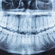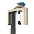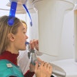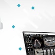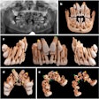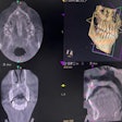
WASHINGTON, DC - 3D cone-beam imaging allows dentists to do and see more, because it offers much greater detail than periapical or panoramic x-rays, according to a well-attended ADA 2015 presentation by dental technology expert John Flucke, DDS.
Benefits of 3D
Although 3D imaging technology shows highly detailed images with greater resolution than traditional imaging technologies, every dentist doesn't need the expensive equipment, noted Dr. Flucke, a practicing dentist in Lees Summit, MO.
 John Flucke, DDS, is a practicing dentist in Lees Summit, MO.
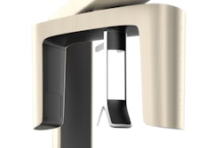
3D technology, also known as 3D cone-beam CT (CBCT), is particularly useful for treatments involving periodontics, endodontics, orthodontics, and oral surgery, he said.
"It depends on how frequently you're doing these procedures, but you get enough information from 3D that we should at least have access to the technology," he told attendees at a session sponsored by the Pride Institute.
Mobile services are available that go to patients’ homes to do scans, as are facilities where patients can go to get them done. Some dental schools will also do 3D scans of patients, Dr. Flucke noted.
Dr. Flucke said he takes images a couple of times per week for specialists, which offsets costs of the system. He takes digital panoramic and bitewing images, then he may take selected periapical images for patients seeking periodontal or endontic treatments. When he has that information, Dr. Flucke decides if a CBCT scan is needed and is worth the extra radiation.
"You've always got to weigh what you need to know versus the risk/benefit to the patient," he stressed. "You only want to use the amount of radiation necessary to get the information you require, so you don't want to take incredibly high-resolution scans in clinical situations where you don't need that much data."
All dentists have had situations in which they would have benefited greatly from cone-beam systems, and everything worked out just fine, Dr. Flucke acknowledged.
"But we owe it to ourselves and our patients to have all the information available," he said, adding that it gives the "big picture" of things. "Often things aren't always the way they appear to be when you look at them on traditional imaging systems, and 3D allows you to see everything."
Why use 3D when 2D works fine?
Dr. Flucke said he's often asked that question by colleagues. He outlined several reasons, including that 3D imaging eliminates surprises during treatment, reducing unanticipated outcomes.
"You don't get in there and find something different than what you thought," he explained. "Also, you don't know what you don't see."
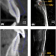
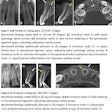
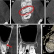
He discussed cases in which mesial buccal roots were split and there were tiny mesial buccal roots, but it wasn't evident on traditional images that these roots were overlapping.
The periapical x-ray won't show the overlapping until you get into the extraction, Dr. Flucke noted, "so sometimes our training sees one thing, but the reality is much, much different."
"I would much rather know about a root that maybe curves away to the palatal or curves into the buccal that you can't see on a radiograph, as opposed to starting the extraction, and ... suddenly then you see the curvature," he said. "I'd rather know about that ahead of time."
Advantages of 3D
Standard digital panoramic x-rays do not show important factors, such as the length of teeth, which cone-beam technology does show, Dr. Flucke said, going on to describe the ability to get the total image of a patient's head.
"I tell patients I can hold your head in the palm of my hand; I can rotate it and look at it and take it apart and look at it from different angles and upside down and see tiny pieces," he said. "Or I can collimate it down if I want to work in one unique area, I can scan just that area and decrease the radiation even further."
“If you're doing something that involves perio or endo and you want to check if there was a canal that was missed, for that you're going to need a high-resolution scan.”
Along with highly detailed resolution, however, Dr. Flucke noted that 3D technology also involves more radiation.
"You'll get more radiation than with digital panoramic x-rays, and that's important to know, because you don't need to shoot everything at high resolution," he said.
And some new systems offer 3D rendering of patients with less radiation than standard digital panoramic x-rays, he said.
If dentist are doing implants and just want to make sure where the alveolar nerve is or if they want to check bone height and the width of the placement of the implant, they can be done with fairly low-resolution systems.
"But if you're doing something that involves perio or endo and you want to check if there was a canal that was missed, for that you're going to need a high-resolution scan," Dr. Flucke pointed out. "You may want to collimate those images down, especially for endo, and just takes images of the teeth on either side of it."
Reading 3D images is not that difficult, he assured the audience.
"I'm not an engineer or a radiologist, just a general dentist," Dr. Flucke said. Describing himself as a "molar mechanic who works in spit," he added, "If I can figure it out, anybody else can too; it's not that difficult."
3D can make a big difference
Dr. Flucke described a case in which a patient complained that a bridge felt loose. Periapical x-rays showed bad margins and problematic endodontics, so he recommended extracting the teeth to support the bridge.
When a 3D scan showed the crown going one way and the root going another way, Dr. Flucke sent the patient to a periodontist for a bone graft.
"But the time to figure this out is not when I'm in the middle of the extraction and thinking we're going to do some easy implants," Dr. Flucke said.
In another case, a woman who had been treated for years in another state came in for standard cleaning, but then 3D imaging showed radiolucency around a tooth.
"I saw this and my blood ran cold," Dr. Flucke recalled.
He sent her to an oral surgeon who operated on her in the hospital, because he was afraid he'd fracture the mandible during treatment. After doing a serious bone graft, she lost a tooth due to the lesion, but the patient healed.
When the patient returned to Dr. Flucke's office, she asked him, "I went to the same office for 10 years, and nobody ever told me anything was wrong. I'm in your office 10 minutes, and you guys find this thing. Why is it nobody else saw it before you did?"
It was a difficult conversation, he said. "I told her I didn't know what it looked like before, only what I saw, and let's just be thankful we saw it before it created problems."
He thinks she had a gag reflex, and the other office probably was trying to be nice when taking x-rays and didn't force the sensor down where it really needed to go to get the image.
"So by trying to be nice to the patient, they actually ended up doing her a disservice, because they didn't get the right image," Dr. Flucke said. "These things are out there, and if I'm seeing them on a regular basis, I know you all are too."
