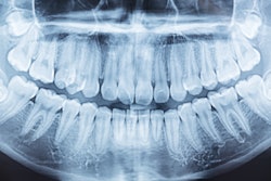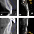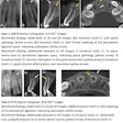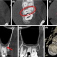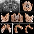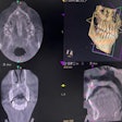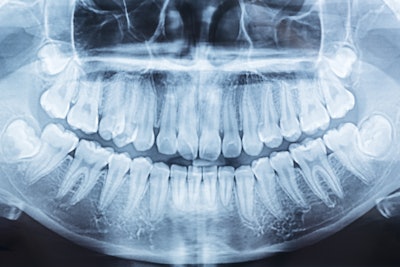
Panoramic (PAN) x-rays performed poorly in detecting external root resorption of third molars, but cone-beam computed tomography (CBCT) did much better in diagnosing this condition. The study was published in October in Clinical Oral Investigations.
Therefore, it may be valuable for patients to undergo CBCT imaging prior to third-molar surgery to avoid removing the teeth without confirming root resorption-induced second-molar damage, the authors wrote.
"Conventional (PAN) imaging is an imprecise method for radiological assessment, with a high frequency of false positive and false negative diagnoses," wrote the authors, led by Dr. Martin Kunkel of the University of Bochum Medical School in Germany (Clin Oral Investig, October 9, 2024, Vol. 28, 583).
Root resorption has been discussed for several decades, but only recently has resorption caused by retained teeth gained attention due to advances in imaging. This subject specifically has been of interest due to the dissemination of quality three-dimensional dental scans.
To compare the sensitivity of x-rays and CBCT in diagnosing root resorption, a retrospective cross-sectional study of 367 patients who underwent both types of imaging was conducted. Past orthodontic treatment, age, gender, superimposition of second molars and third molars on x-rays, retention depth, inclination angle, and vertical level of contact with second molars were used as predictor variables, according to the study.
Less than 5% of panoramic x-rays revealed resorption linked to third molars, and CBCT showed resorption in 20% of second molars with adjacent retained third molars. Predictive parameters for restoration were found to be the angle of inclination of third molars, age, and vertical level of molar contact. Additionally, increased severity of second molars was linked to mesial inclination, older age, and deeper retention, the authors wrote.
Furthermore, about 16% of second molars showed root resorption on CBCT despite showing superimposition on panoramic x-rays. Therefore, conducting CBCT imaging before third molar surgery may be beneficial to prevent removing the teeth without knowing about second molar damage due to resorption, they wrote.
Nevertheless, the study had limitations. Due to the accumulation of dependent observations, using third molars as the type of tooth in this research may lead to an overestimation of possible risk factors, the authors wrote.
"Taken together, our results confirm that RR is not a rare phenomenon, and that PAN-imaging alone is not sufficient to depict RR," Kunkel and colleagues wrote.


