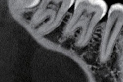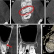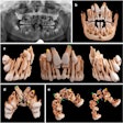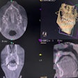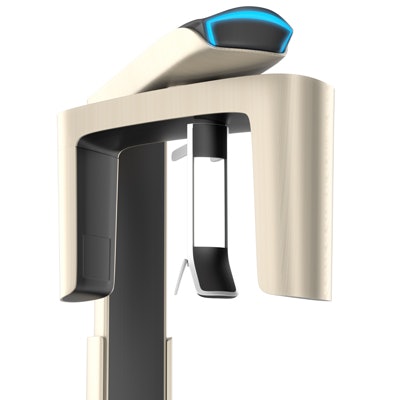
Cone-beam CT (CBCT) shows promise for periodontal diagnosis, but how does its use measure up against conventional imaging? To learn more, researchers compared the results of studies on the use of CBCT for assessing periodontal conditions and parameters.
They searched for studies that compared CBCT with conventional imaging techniques for assessing periodontal parameters, including infrabony defects and furcation involvement. Their review findings indicated that CBCT was superior.
"CBCT is significantly more accurate and reliable than two-dimensional conventional imaging techniques for assessing infrabony defects, furcation lesions, the height of the alveolar bone crest, and the periodontal ligament space," the authors wrote (Imaging Science in Dentistry, June 2018, Vol. 48:2, pp. 79-86). "However, differences in imaging protocol parameters can affect the reproducibility and reliability of CBCT measurements."
The lead author was Isabela Goulart Gil Choi of the oral radiology department at the University of São Paulo School of Dentistry in Brazil.
Image management
The use of CBCT for periodontal diagnosis demonstrates potential benefits, although drawbacks include both greater expense and larger radiation dose compared with conventional imaging techniques, the authors noted. However, knowledge is limited regarding how conventional and 3D imaging compare for diagnostic performance, precision, and accuracy.
Therefore, the researchers conducted a systematic review of studies that directly compared the precision and accuracy of CBCT with conventional imaging techniques for assessing the following periodontal conditions and parameters:
- Infrabony defects
- Furcation involvement
- Alveolar bone crest height
- Periodontal ligament space
The researchers searched the Medline and Embase databases for original articles, systematic reviews, and case reports published in English through 2017. They required that included studies reported the advantages and disadvantages of visualizing some of these periodontal conditions and parameters with more than one diagnostic imaging technique.
“Differences in imaging protocol parameters can affect the reproducibility and reliability of CBCT measurements”
The review included 13 studies, of which four discussed infrabony defects, three examined furcation involvement, three looked at the height of the alveolar bone crest, two studied periodontal ligament space, and one evaluated infrabony defects, furcation involvement, and periodontal ligament space. Only four of the studies were conducted in vivo, and they used different scanning parameters, which made direct comparisons difficult, the authors wrote.
Two of the four studies on infrabony defects dealt with naturally occurring defects in human skulls, and the other two evaluated simulated infrabony defects. The studies on artificially created infrabony defects compared measurements made with periapical radiographs and CBCT with control measurements directly made on dry skulls. The authors of these studies reported that CBCT produced greater precision and accuracy than the periapical radiographs because of CBCT's advantage of identifying defects in both the buccal and lingual aspects of teeth.
The one study that looked at the accuracy of CBCT for identifying clinical bone defects found that CBCT produced more false positives when detecting fenestrations compared with direct visualization.
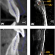
One study compared measurements of periodontal defects made with periapical radiographs, panoramic radiographs, CT, and CBCT with histological specimens and found better image quality with CBCT and also smaller deviations in periodontal defect dimensions than with histological results.
Of the three studies on alveolar bone crest height, one examined defects in human skulls, one evaluated correlations in vivo between CBCT and direct surgical measurements after bone replacement graft procedures, and one analyzed images from a database of patients referred for periodontal evaluation. The studies found that CBCT was more accurate than conventional radiographs for assessing alveolar crest height due to less underestimation of horizontal bone loss.
Among the studies on furcation involvement, two used intraoperative findings as the gold standard and found that CBCT and surgical measurements agreed in 84% of cases. A third study was conducted on human skulls and found CBCT images were more accurate than intraoral digital radiography at identifying furcation defects. The fourth study examined simulated lesions in macerated pig mandibles and found that CBCT images were highly accurate (78% to 88%) at detecting furcation involvement.
The studies on furcation involvement also found that CBCT provided important information about root morphology and residual attachment of maxillary molars, a significant advantage compared with conventional presurgical clinical assessment methods.
Of the remaining studies, one was a human ex vivo investigation that confirmed close agreement between histological findings and CBCT for infrabony defects, furcation involvement, and periodontal ligament space. The results of the few studies that examined the accuracy of imaging for evaluating the periodontal ligament space have been mixed, with some having found CBCT to be more accurate than conventional radiography at detecting marginal widening.
Periodontal assessment
The authors noted some limitations of their study:
- Most articles reviewed were based on in vitro experiments.
- Study observers had varying levels of expertise with CBCT.
- There was a lack of studies that qualitatively or quantitatively compared conventional imaging techniques with CBCT.
Nonetheless, they determined that CBCT performed better for assessing periodontal issues.
"These studies revealed CBCT to be the best imaging technique to assess infrabony defects, furcation lesions, the height of the alveolar bone crest, and the periodontal ligament space," the authors concluded.
