Pulp stones are nodular, calcified masses that may appear in the coronal and root portions of a tooth's pulp organ. When it comes to identifying these masses, is cone-beam CT (CBCT) or digital panoramic radiography more effective? Researchers compared the two modalities to find out.
They looked at both CBCT and digital panoramic images for more than 200 patients and found that CBCT was the better option for finding these masses. They published their study in Imaging Science in Dentistry (September 2018, Vol. 48:3, pp. 201-212).
As a 2D imaging system, digital panoramic radiography "has inherent limitations leading to the misinterpretation of pulp stones," wrote the authors, led by Melek Tassoker an assistant professor in the department of oral and maxillofacial radiology at the Necmettin Erbakan University Faculty of Dentistry in Konya, Turkey.
Age correlation
The prevalence of pulp stones varies widely in published studies, from less than 10% to more than 90%. As previous literature has suggested that 2D radiographic techniques may underreport such calcifications, the researchers wanted to compare CBCT and digital panoramic radiography for detecting these masses and also identify any potential correlation between the presence of pulp stones and factors such as age, sex, the tooth involved, restoration, and caries.
The researchers randomly selected both digital panoramic and CBCT images of 202 patients from their department's database. They recorded the systemic condition of patients, presence of pulp stones, location of the tooth, group of teeth, and presence and depth of caries and restorations. For reasons related to image quality, they compared only the images of posterior teeth (1,616 molar teeth) in terms of pulp stone occurrence.
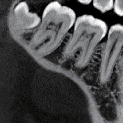
 Left: Cropped panoramic radiograph shows pulp stones in the upper and lower molars. Right: Coronal CBCT slice shows pulpal calcifications in the lower molars. Images courtesy of Imaging Science in Dentistry (September 2018, Vol. 48:3, pp. 201-212).
The team identified 105 pulp stones in the 202 subjects. The prevalence of pulp stones was similar between the sexes and across various tooth locations and groups of teeth (p > 0.05). Overall, both of the modalities found 152 of the 1,616 teeth had pulp stones, whereas digital panoramic radiography alone detected 252 pulp stones.
“CBCT is a superior method for detecting pulp stones compared to conventional radiography.”
However, calcified masses smaller than 200 µm cannot be seen on 2D radiographs, making difficult it for digital panoramic radiography to distinguish such small calcifications from the results of overlapping with other tissues, the study authors noted.
"CBCT is a superior method for detecting pulp stones compared to conventional radiography, as it has greater specificity and accuracy and overcomes the overlapping limitation," they wrote.
The researchers also reported that maxillary canine teeth had the highest occurrence of pulp stones (9.7%), followed by mandibular incisors (9.2%), mandibular premolars (8.3%), and mandibular canines (6.4%).
In addition, teeth with restorations and caries showed a significantly higher prevalence (73.5% and 62.1%, respectively) of pulp stones than teeth without restorations or caries (p < 0.05).
Better for identification
The authors noted that this was a cross-sectional study and suggested that longitudinal studies with larger sample sizes should be conducted.
Because of the limitations of 2D digital panoramic radiography, the authors recommended using CBCT to identify these masses if they are suspected on digital radiography.
"CBCT imaging is advised for the examination of pulp stones once there is a suspicion of a pulp stone on [digital panoramic radiography]," they concluded.

