DrBicuspid.com is pleased to present the next installment of Leaders in Dentistry, a series of interviews with researchers, practitioners, and opinion leaders who are influencing the practice of dentistry.
We spoke with Alan Rosenfeld, DDS, and George Mandelaris, DDS, MS, periodontists who have been practicing together in the Chicago area since 2000. Their ground-breaking work with Richard Mecall, DDS, set the educational and professional standard for the use of CT scans for diagnostic purposes, surgical treatment planning, computer-guided implant placement, and determination of prosthetic outcomes in the dental profession.
Drs. Rosenfeld and Mandelaris shared their thoughts on the evolving role of periodontists in interdisciplinary dentistry and their recent experiences with cone-beam CT in their practice, which they believe will open up new diagnostic and treatment opportunities for periodontists.
DrBicuspid: How long have you been working with 3D imaging in your practice?
 Alan Rosenfeld, DDS
Dr. Rosenfeld and Dr. Mandelaris: Since 1988. Our initial foray into the use of medical-grade CT imaging was strictly hospital-based, but this was very inconvenient for our patients to be imaged on a timely basis. That changed over time to being able to obtain CT images from imaging centers.
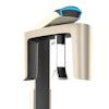
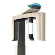
The Carestream 9300 is our first involvement with cone-beam CT on a regular basis. We have been working with it since June and are the first practice in the U.S. to have it. We decided to work with this machine because it offered the greatest variation of imaging possibilities for our patients. Depending on our diagnostic inquiry, we can limit exposure to specific areas of concern for the patient. For a long time we were caught in a quandary between the high quality of images we see with medical-grade CT and what we can obtain with cone-beam CT. So the ability to get equivalent images with substantial reduction in radiation exposure to patients was a primary motivator to adopt the cone-beam CT technology.
What new capabilities does cone-beam CT bring to your practice?
 George Mandelaris, DDS, MS
Dr. Rosenfeld: In addition to reducing radiation exposure, we now also get to control the quality of the imaging. We use a radiology technologist to provide our images -- the same person, not a different technician depending on the day or schedule. And we are working with a person who was involved with providing us the medical-grade images from an imaging center. So he has a tremendous amount of experience.
Dr. Mandelaris: The 9300 provides high-quality cone-beam CT images with individualized field-of-view exposures and significantly lower radiation exposure, especially compared with medical-grade CT. So we're not compromising the diagnostic capabilities we've become accustomed to. This means we can use it on a more routine basis without exposing patients to radiation beyond what they would typically get with a full-mouth radiographic exam using D-speed film.
Dr. Rosenfeld: When we were dealing with higher radiation doses, such as medical-grade CT scans, we were largely doing referrals to imaging centers for implants. Not for oral pathology related issues, just implants. But now, the ability to incorporate cone-beam CT allows us to use this technology to support other things in periodontics that we think are part of a paradigm shift. All the things we now do using computers -- electronic records, digital x-rays, 3D imaging -- it is all information management. These technologies are all extensions of our ability to manage information and get instantaneous access to information that helps with patients and strengthens our relationships with referral doctors.
What kinds of periodontal applications are you now using cone-beam CT for?
Dr. Mandelaris: Cone-beam CT doesn't have to be a tool that is just used for implant diagnostics any longer. And this is partly because of the robust viewing software, which allows you to look at so many different angles and positions of the teeth. We've been using it for implant diagnostics and some periodontal disease diagnostics, and also endodontic considerations and dentoalveolar applications. You can use it to evaluate endodontic pathology, do temporomandibular joint assessment, analyze airway issues for sleep apnea patients, evaluate pathology fractures, and do trauma-related 3D analyses for oral and maxillofacial services. You can order a single arch, a few teeth, or an entire maxillofacial complex.
Dr. Rosenfeld: People are pretty clear about the use of this technology for implants, but they may not be clear about one of the significant emerging opportunities: surgically facilitated orthodontic therapy. That is, creating an accelerated rate at which teeth can be moved in a more efficient and safe manner. But to properly diagnose these patients -- most have bone and gum tissue deficiencies, which can cause complications. So with cone-beam CT, we can evaluate the tooth-bone-gingival complex, analyze the whole thing. Most people don't see recession as an issue, but recession is actually bone loss. And what you see in the mouth is actually a small amount of what is really there.
How can this technology change the way periodontists diagnose and treat?
Dr. Rosenfeld: What is really fabulous about this technology is, if you have the question, you just need to manipulate the software to get your answers. What is rich about the 9300 is that you can manipulate the data so you can see things you can't typically see on other kinds of imaging software. For example, you can adjust the information so you can look exactly down the long axis of the tooth and analyze it that way.
Cone-beam CT is really a convenient adjunct to things we are already doing in dentistry. You can look for endodontic problems and fractured teeth we couldn't see before. We're just beginning to learn about the possibilities. I don't think cone-beam CT replaces original forms of clinical assessment. But radiation exposure to get certain kinds of information is unnecessary. When you have a particular periodontal issue and you can't get the answer using traditional means, you can use cone-beam CT and zero in on the particular teeth you are trying to inquire about, versus doing a full-face or medical-grade CT.
Dr. Mandelaris: Periodontics is the most interwoven discipline when it comes to being involved in general dentistry and being a partner with general dentists to support prosthetic needs. We are involved with implants, we manage dentoalveolar structures and inflammatory periodontal diseases, recession-based attachment loss, perio-restorative, and we provide a multitude of plastic and reconstructive procedures for orthodontic applications. We are not just talking about gum disease.
To be able to assign prognoses to teeth, you need a full understanding of regional and local anatomy. So, in addition to using our clinical exam and validating these decisions with evidence-based research, we now have a more informative tool. With 3D assessment, you have the ability to look at all the nuances, furcation involvements, and the extent and location of remaining bone in much greater detail. It puts periodontists in an even more critical position to enable correct diagnoses and assign more accurate prognoses. This can only help to develop more effective treatment plans with our interdisciplinary teams. If you have more information and all the cards are on the table, you can make better treatment decisions, which is obviously in the best interest of the patient.
What will it take for more periodontists to see the value in adopting this technology into their practices?
Dr. Rosenfeld: I'm 62, and in my age group, unless you are still passionately engaged, you're probably not going to get involved with this technology unless you're bringing in an associate who has the energy and wherewithal to help introduce this into a private practice setting. Because once you get involved in cone beam, you also have to have the infrastructure in your office to support information acquisition.
But the 35- to 52-year-olds, the people who are in the middle of their professional careers -- it is going to be incumbent upon them to make this transition because that is where the profession is going and it is what is expected. Cone-beam CT can help make the judgment that teeth are savable, and we are the only profession in dentistry that is focused on saving teeth. This is where most periodontists will be moving to in the future. If you can diagnosis better, ask better questions, and be better prepared for treatment, this technology is a gift.
Dr. Mandelaris: There are three stages of a scientific theory. In the first stage, the theory is scoffed at and met with disbelief. In the second stage, it is accepted as true but is considered insignificant and trivial. In stage three, it is thought to be correct and even revolutionary. In fact, those who criticized it most now claim that they invented it and are the experts.
CT-based decision-making has been a 25-year journey in our practice but has seemingly become an overnight phenomenon in dentistry. The opportunity that cone-beam CT imaging presents to contemporary periodontists is incredible. This opportunity demands our involvement in interdisciplinary dentistry. By having a better prognosis, you can change the course of someone's complex treatment and perhaps effect change in their life.
The pendulum has swung so far to implants that saving natural teeth has become too casual of a conversation. If we have more information to make better treatment decisions and use evidence-based literature to make these treatment decisions, saving more teeth becomes a more important and realistic treatment option for patients and one that provides a better-informed consent before irreversible procedures are performed.
So in our practice, it's not about cone-beam CT, it's about information management and making good treatment decisions with that information.
