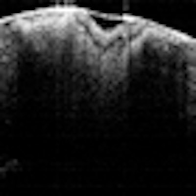
The ability of optical coherence tomography (OCT) to image the progression of caries beneath dental sealants could be the catalyst that helps practitioners see the clinical value of this diagnostic tool, according to one public health advocate.
A new study in Lasers in Surgery and Medicine (September 16, 2010) "has advanced this concept one stage closer to the clinical situation by identifying the diagnostic usefulness of OCT images within a simulated clinical setting -- that is, the ease with which clinicians can detect demineralization (early lesion) on the tooth surface underneath resin or GIC [glass-ionomer cement] sealants from a rapid examination of an image -- thus corresponding to current usage with radiographs."
“OCT is really the only way to look at infinitely small changes in enamel or dentin.”
— Jennifer Holtzman, DDS, MPH,
Beckman Laser Institute
Current technology does not permit monitoring of potential lesion progression or arrest, noted Jennifer Holtzman, DDS, MPH, lead author on the study, which was conducted in conjunction with colleagues from the University of California, Irvine and University of Pennsylvania School of Dental Medicine. This can prompt some clinicians to opt to place restorations instead of dental sealants, she told DrBicuspid.com.
"One of the issues in dental public health is the reluctance of some practitioners to place dental sealants," said Dr. Holtzman, who as a faculty member at the University of Southern California ran the school's community-based dental sealant program for nine years before recently becoming an assistant researcher at the Beckman Laser Institute. "Their issue is 'if I place dental sealant on a tooth that may have some caries, how do I know it is arrested under this sealant and not progressing?' So we wanted to find out if there was a way to see caries and caries progression underneath the sealant."
Four common sealants
For this study, she and her colleagues divided 40 extracted teeth into equal groups of carious and noncarious teeth (based on visual inspection). They were considered carious if there were white or brown spot lesions on the tooth not consistent with the clinical appearance of sound enamel.
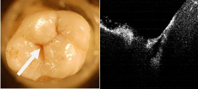 |
| Left image: Molar with early occlusal caries; arrow points to area imaged at right. Right image: OCT of pit. |
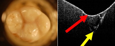 |
| Left image: Same tooth with sealant. Right image: OCT of same area postsealant. Red arrow points to sealed surface; yellow arrow points to decay. |
The teeth were imaged using standard radiography and swept-source spectral-domain OCT (1350-nm wavelength), then randomly assigned for sealant placement using one of four commonly used dental sealants: Clinpro (3M ESPE), Fuji Triage (GC America), Embrace Wet Bond (Pulpdent), and Delton (Dentsply). Following sealant placement, the teeth were imaged again with OCT and radiography, in the same locations as the presealant images.
|
Types of OCT Several types of OCT are being developed and investigated for use in dentistry, including time-domain OCT, polarization-sensitive OCT (PS-OCT), and spectral-domain OCT (SD-OCT), although there are very few commercial products available. Each type of OCT has its pros and cons; for this study, the researchers chose a swept-source SD-OCT system to take advantages of the faster imaging time and higher sensitivity and demonstrate the practical side of this technology. "Because our goal was to determine the clinical feasibility and utility of the proposed approach, fast and simple visual examination of OCT images by clinicians such as would be convenient at the chairside was used for the imaging-based diagnosis," the researchers wrote. Other groups are studying PS-OCT for diagnostic applications in dentistry. Daniel Fried and colleagues at the University of California, San Francisco have published a number of papers looking at the ability of PS-OCT to image demineralization and remineralization of caries lesions in the dentin, including under sealants (Journal of Biomedical Optics, November 2004, Vol. 9:6, p. 1297). "One neat thing about PS-OCT is that the sealant becomes invisible in the cross-polarization images, so you can quantify the decay under the sealant or composite," he told DrBicuspid.com. |
The researchers found that the OCT imaging was rapid and simple, with an average imaging time of less than 1 minute per tooth, and tooth structures were easily identifiable using visual evaluation.
"After 90 minutes of training, prestandardized dentists were able to detect tooth decay more accurately using OCT than with visual or radiographic examination," Dr. Holtzman and her colleagues wrote.
Caries/demineralization was easily recognized in the OCT images by its differing optical intensity from the adjacent tooth substance and sealant, they noted. Underneath sealants, areas of demineralization/decay were visible as very bright zones that could be clearly distinguished from the overlying sealant.
Diagnostically, visual exam falsely identified six decayed teeth as being healthy. Prior to the sealant application, the radiographs missed early lesions in 20 specimens, while OCT misdiagnosed two healthy specimens as being decayed. Following sealant application, radiographs missed early lesions in 15 specimens, while only three lesions went undetected using OCT.
As evaluated using positive predictive values and negative predictive values, the detection of demineralization or caries was more accurate with OCT than with visual or radiograph examination, the researchers noted. Of the four dental sealants, they found that Delton provided "excellent positive predictive values and the best postsealant negative predictive values."
'A huge bargain'
Despite the diagnostic advantages OCT demonstrated in this study, barriers to adoption remain. There are very few commercial OCT systems designed specifically for dental applications -- Lantis Laser has done the most development work in this area, although Panasonic Shikoku Electronics reportedly is developing a dental-specific OCT system and Michelson Diagnostics is collaborating with researchers at Kings College London to evaluate the use of its OCT system for assessing dental implant failures -- so availability is an issue. But Dr. Holtzman and her colleagues are convinced that the technology presents a tremendous opportunity to improve diagnostics in the oral cavity.
"Clearly, clinicians' concerns regarding their current inability to detect demineralization are justified," the researchers wrote. "The decision to restore teeth rather than let demineralization potentially progress unmonitored underneath sealants is understandable, yet it is not optimal. The development and validation of an improved tool for monitoring demineralization status under dental sealants may well increase dental sealant utilization, especially in high-risk and underserved populations."
In addition, providing clinicians with a tool such as OCT that can reliably confirm the presence of sound enamel would reduce the number of unnecessary restorations or sealant replacements, the authors noted, resulting in better utilization of manpower and time and reduced healthcare dollars.
"If you look at how healthcare is slowly changing from a surgical model to a preventive model, OCT is really the only way to look at infinitely small changes in enamel or dentin," she said. "So once dentistry really goes from that surgical model to a wellness model, I think OCT will catch on."
While the cost of an OCT system -- typically around $60,000, according to Dr. Holtzman -- may still give some practitioners pause, she considers OCT a "huge bargain" in terms of its ability to help practitioners feel more confident about dental sealants and whether the caries is progressing.
"For the dentist, the risk of being wrong, of missing the caries -- that is something none of us wants," she said.
Copyright © 2010 DrBicuspid.com


