Can the accuracy of immediate implant placement be improved in patients with terminal dentition by using tooth-supported, bone-reduction guides?
In a study published in the Journal of Oral Implantology (June 8, 2016), researchers from San Francisco and Kuwait found that CAD/CAM surgical guides are essential for implant placement but wondered if current methods could be improved. They specifically tested the "three-guide technique."
"CAD/CAM surgical guide fabrication is an emerging tool that may facilitate the surgical process and aid in safe and predictable execution of bone reduction and immediate implant placement," the authors wrote.
Terminal dentition
Patients with terminal dentition who need immediate implants can be a clinical challenge, especially if alveoloplasty is necessary to make interarch space for the implants, the authors wrote. For these patients, CAD/CAM surgical guides are essential. However, existing techniques, whether mucosa or bone-supported, often don't have enough guide stability and accuracy, even with anchor pins, compared with teeth-supported guides, they noted.
This is the first study to investigate the use of CAD/CAM technology for bone reduction before implant placement in these completely edentulous patients and/or patients with terminal dentition, according to the authors.
The study included five patients (two women and three men): Three patients had terminal dentition in their mandibular arches, and the other two had maxillary terminal dentition.
After obtaining images, the researchers studied the surgical guides thoroughly before beginning the implant procedures. For each procedure, the patients were sedated and the first CAD/CAM guide was placed. Surgical drills were used to create 2.0-mm osteotomies through the soft tissue to place the anchor pins as directed by the guides. Three anchor holes were used to fix the location of all subsequent guides after extraction. Postsurgical images also were taken.
In total, the researchers placed 26 implants in five arches. All implants placed were NobelReplace Conical Connection (Nobel Biocare).
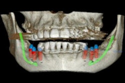
To evaluate the accuracy of the three-guide technique, the researchers evaluated both pre- and postsurgical cone-beam CT (CBCT) images of the bone reductions and the implant placements with the simulated and the actual implants superimposed. Three sites were selected on each arch (right, middle, and left). They measured deviations from the surgical plan at three points, which were then averaged and presented as a single deviation at the site. Total deviation was then calculated based on the averages of the three sites (see table below).
For the five patients, the overall average deviation of the implants at the crest was 1.43 mm and 1.9 mm at the apex, with an angular deviation of 4.14°.
| Bone reduction deviations between planned & actual bone reduction | ||||
| Patient number | Right deviation (mm) | Middle deviation (mm) | Left deviation (mm) | Overall average deviation |
| 1 | 1.69 | 2.42 | 2.55 | 2.22 |
| 2 | 1.95 | 2.54 | 2.06 | 2.18 |
| 3 | 1.10 | 1.20 | 0.95 | 1.08 |
| 4 | 2.31 | 2.55 | 2.32 | 2.39 |
| 5 | 1.84 | 2.68 | 1.56 | 2.03 |
| Overall average | 1.78 | 2.28 | 1.89 | 1.98 |
More predictable outcome
The authors emphasized that these patients' bone quantity and quality are reduced due to the removal of the cortical layer. They noted that the three-guide technique offers a "more predictable way" to place the implants in their desired and most optimal position based on the bone quality noted on the presurgical CBCT images.
"This method may improve guide stability for patients with terminal dentition undergoing complete implant-supported treatment by taking advantage of the teeth to be extracted," the authors wrote.
