How well does your root canal sealer do its job? Researchers compared the 3D microporosities and sealing abilities of two sealers, one relatively new and one considered the gold standard. While both performed well, neither achieved a porosity-free filling.
Researchers investigated the effectiveness of sealers AH Plus (Dentsply DeTrey) and the newer EndoSequence BC (Brasseler) at closing dental tubules of human premolars in the laboratory. Both sealers performed similarly, with neither achieving a porosity-free filling and closure levels differing by section.
"A similar volume of closed pores was observed between the EndoSequence BC sealer and the AH Plus, which indicated that tested sealers adapted or penetrated equally to the dentin in the coronal, middle, and apical sections," the authors wrote (Journal of Applied Oral Science, January 15, 2018).
The lead author was Yan Huang, PhD, of the State Key Laboratory of Oral Diseases and the National Clinical Research Center for Oral Diseases at Sichuan University in Chengdu, China, and also the University of Leuven Faculty of Medicine in Belgium.
Sealing it up
Long-term endodontic treatment success requires complete filling after root canal obturation, the authors noted. Treatments may fail for many reasons, including poor contacts between the gutta-percha and sealer or the sealer and dentin, as well as through openings within the sealer.
Using an endodontic sealer along with gutta-percha is helpful because achieving complete filling with root-filling materials is difficult, the authors explained. Adaptability of a sealer to dentin is key.
In the current study, the researchers sought to evaluate the sealing ability of EndoSequence BC sealer, a bioactive product introduced around 2013 that contains nanoparticles about 2 µm in diameter.
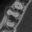
They used scanning electron microscopy and microcomputed tomography (micro-CT) to quantitatively measure the effectiveness at sealing apical, middle, and coronal dental tubules of this product and compared it with AH Plus, previously reported to be the gold standard. No previous studies have used these technologies for this application, the authors noted. Micro-CT can scan filled roots and reconstruct them three dimensionally to assess a sealant's adaptation to root canal walls, they explained.
The researchers conducted the study in 24 similarly sized, single-rooted, human mandibular premolars extracted for orthodontic reasons. They began the study thinking there would be no difference between the two sealers.
They adjusted each root to about 12 mm in length, then randomly assigned each tooth to one of two groups (n = 12). They filled each tooth with a single cone of 40/06 gutta-percha and one of the two sealers, following the manufacturer's instructions and ensuring even distribution.
The researchers divided the roots into three sections: apical (0 mm to 4 mm), middle (4 mm to 8 mm), and coronal (8 mm to 12 mm). They then conducted both a microporosity analysis on a 2-mm-diameter fixed ring area and a scanning electron microscopy analysis.
They performed 3D imaging analysis for each section to calculate the porosity of the sealant, volume of closed pores, surface of closed pores, volume of open pores, and open porosity percentage. Closed pores were dentin tubules already filled or sealed.
Both sealers performed sufficiently along the whole length of the root canal, with better sealing measured in the coronal than apical sections of the molars, according to the scanning electron microscopy results.
The volume and surface of closed pores were the largest in the coronal sections, followed by the middle and apical sections for both sealants (p < 0.05). In all three sections, the researchers found no significant differences for the volume of closed pores and the surface of closed pores between the sealers, although they were larger in the apical section with the AH Plus sealer. These results demonstrated that the two sealers penetrated equally to the dentin in all three sections, the authors noted.
For both sealants, the volume of open pores was significantly larger in the coronal section than in the apical section (p < 0.05). In all sections, the EndoSequence BC sealer had a larger volume of open pores than the AH Plus sealer (p < 0.05) (see table below).
| 3D structural parameters as measured by micro-CT | ||||||
| Apical third | Middle third | Coronal third | ||||
| AH Plus sealer | BC sealer | AH Plus sealer | BC sealer | AH Plus sealer | BC sealer | |
| Volume of closed pores (mm3) | 0.151 | 0.115 | 0.354 | 0.408 | 0.620 | 0.861 |
| Surface of closed pores (mm2) | 0.231 | 0.189 | 0.322 | 0.431 | 0.636 | 0.763 |
| Volume of open pores (mm3) | 4.49 | 6.24 | 6.10 | 6.84 | 6.32 | 6.72 |
| Open porosity percentage | 64.24% | 73.86% | 70.24% | 83.25% | 75.39% | 82.74% |
More research needed
The authors noted that the natural acidity of AH Plus may limit its bonding to dentin and that it contains a polymer that contracts upon polymerization, which could lead to sealant cracking and deterioration.
"It is likely for these reasons that the EndoSequence BC sealer shows superior sealing ability than AH Plus, but [this] has yet to be confirmed by further in vivo follow-up studies," they wrote.
But for now, the researchers did not find any differences in the performance of the sealers.
"By using the single-cone technique, neither EndoSequence BC or AH Plus provides a porosity-free root canal filling," the authors concluded. "The EndoSequence BC may have similar sealing abilities regarding the whole root canal as the AH Plus sealer. A better sealing effect could be obtained in the coronal and middle sections of a root canal than the apical part by using the tested sealers."


