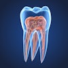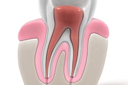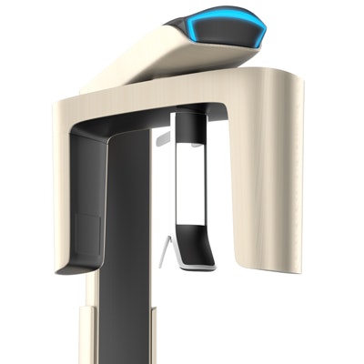
The first step in planning for root canal treatment is to be able to clearly and accurately visualize the anatomy of the canal. European researchers wanted to know if a cone-beam CT (CBCT) system can be used to help develop a treatment plan.
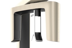
Testing two devices, the Scanora 3D (KaVo) and the 3D Accuitomo 170 (J. Morita Europe), the researchers examined scores of images from each system and found that they were equally acceptable for use in endodontic treatment planning.
"Both systems seem to be equally useful in endodontics, which is a very interesting finding regarding other CBCT-related factors such as installation and maintenance costs, possible field-of-view and range of application, and additional options," the authors wrote (Swiss Dental Journal, March 2017, Vol. 127:3, pp. 221-229).
The study was led by Dr. Sabrina Strobel of the department of operative dentistry and periodontology at University Medical Center Freiburg in Germany.
Accuracy is crucial
The success of endodontic diagnosis and treatment often depends on being able to accurately view the root canal's anatomy. The researchers wanted to learn if a CBCT scan could be used to detect vertical root fractures, number of roots, lateral canals, and the presence of fillings or posts to help plan endodontic treatment.
For their in vitro study, they used 70 extracted human teeth, single- and multirooted teeth and also teeth with and without previous endodontic treatment. The team randomly numbered and scanned the teeth with a digital imaging plate system (Gendex Oralix AC, KaVo) before imaging them with the two CBCT systems.
“Both systems seem to be equally useful in endodontics, which is a very interesting finding regarding other CBCT-related factors such as installation and maintenance costs, possible field-of-view and range of application, and additional options.”
The researchers asked the following questions:
- What percentage of detectable roots, root canals, and lateral canals were seen?
- What percentage of root canal fillings and posts were seen?
- Was the presence of vertical fractures detected?
- Did artifacts appear?
After the CBCT scans, the researchers photographed the extracted teeth and then cut them into cross sections to be used as a gold standard for the comparison and evaluation. A study coordinator compared the cross sections with the evaluated 3D results of the CBCT systems. The cross sections showed a total of 129 roots, 185 root canals, 81 natural lateral canals, 86 roots with root fillings, 8 posts, and 12 vertical fractures within the 70 teeth.
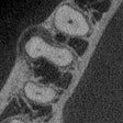
Two general dentists evaluated two 3D CBCT datasets of every tooth in all possible views (axial, coronal, and sagittal). Both CBCT systems were acceptable for use in endodontic treatment planning based on the percentage of correct diagnoses rendered with each product, the authors reported. What's more, they found no statistically significant differences between the two systems.
The results are shown in the table below.
| Summary of correct diagnoses by CBCT system | |||
| Criteria | No. | 3D Acuitomo 170 | Scanora 3D |
| Roots | 129 | 97.2% | 98.6% |
| Root canals | 185 | 100% | 98.6% |
| Lateral canals | 81 | 100% | 98.6% |
| Root fillings | 86 | 100% | 92.9% |
| Posts | 8 | 95.8% | 95.8% |
| Vertical fractures | 12 | 100% | 98.6% |
| Enamel-dentin differentiation | N/A | 78.6% | 61.4% |
| False diagnoses | 24/140 | 9/140 | |
The authors also reported that artifacts were found in almost 90% of CBCT images across both systems, with the artifacts labeled more distinctive by the Scanora 3D. However, this is a subjective conclusion, they noted. They also labeled so-called "beam-hardening artifacts" (caused by any high-density material within the scanned area) as being more severe in the images taken from the Scanora 3D.
Visualizing complex structures
The study authors noted that the CBCT systems tested are different from each other and that there were no specific guidelines for adjustments, leading them to try to keep adjustments within manufacturer's recommendations on voxel size and field-of-view. They recommended further study, with a larger number of participating dentists and using radiologists to make adjustments to the devices.
In addition, the authors noted that both dentists who evaluated the CBCT images detected a post within the 3D dataset of one tooth, although there was none actually. This could be caused by the root canal anatomy (or a previous existing but removed post) or perhaps a very thick and well-condensed root filling, they speculated.
However, both CBCT systems are convenient and well-suited for visualizing complex endodontic structures, the authors wrote.
"Both CBCT systems were found to be similarly suitable for the visualization of endodontic structures in vitro," they concluded.

