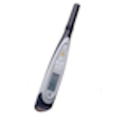
Think the Diagnodent is only good for helping to confirm the presence of caries or calculus? Think again. A new study in the Journal of Endodontics (October 2009, Vol. 35, pp.1404-1407) has found that laser fluorescence can also enhance the assessment of endodontic infection.
Evaluating the microbial status of the root canal system typically involves taking a culture. But the drawback with cultures is that they take time to grow, and they only assess the status of the tooth at the time the sample is taken, said John M. Nusstein, D.D.S., M.S., director of the Advanced Endodontic Clinic at the Ohio State University College of Dentistry and chair of the American Academy of Endodontics’ Research and Scientific Affairs Committee.
"When one re-enters the tooth several days later, the actual microbial status may be completely different," he said. In addition, not all areas of the root canal can be sampled, nor can specific areas be targeted for sampling.
Sapphire tips
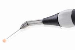
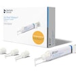
These limitations prompted researchers from the University of Queensland School of Dentistry to try a different approach. First, they fitted a Diganodent Classic with a sapphire tip that has the same design and optical properties as the Diagnodent Pen's periodontal "P" tip. They then used the device's visible laser light (655 nanometers, red) to elicit near-infrared fluorescence emissions from infected and uninfected root canals, enabling them to evaluate the fluorescence properties of bacterial cultures, monospecies biofilms in root canals, pulpal soft tissues, and sound dentin.
Fifty unerupted, extracted third molars were prepared using rotary nickel-titanium instrumentation and EDTA and sodium hypochlorite as irrigants, then autoclaved and inoculated with either Streptococcus mutans or Enterococcus faecalis, whose fluorescence emissions are less intense than those of gram-negative bacteria. The teeth were cultured at 37 degrees Centigrade for seven days under enriched carbon dioxide conditions to allow a biofilm to develop in each root canal. Fluorescence readings of the canals were taken after three and seven days.
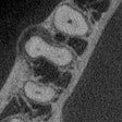
To obtain the fluorescence readings, the researchers placed the Diagnodent tip in direct contact with the soft tissue of the dental pulp. The pulp was then removed and readings taken of each canal orifice. Next, the pulp tissues were extirpated and the probe immediately reinserted into the root canal for further fluorescence measurements. To confirm the presence of biofilms, 10 specimens were split and examined using scanning electron microscopy (SEM).
A second part of the study focused on 50 teeth that had been extracted due to pulpal pathosis. Each had radiographic and clinical evidence of pulp death in the form of periapical pathology and frank carious exposure. After sterilizing the teeth, the crowns were removed and the Diagnodent probe inserted, with fluorescence measurements taken from the deepest insertion into the canals. Roots from 20 of these teeth were then examined using SEM to confirm the presence of infection.
Fluorescence findings
In reviewing the SEM scans, the researchers found that sound dentin and healthy pulpal soft tissue gave an average fluorescence reading of 5 out of a scale of 100, whereas biofilms of Enterococcus faecalis and Streptococcus mutans established in root canals showed a progressive increase in fluorescence over time. Fluorescence readings fell to the "healthy" threshold (5) when the root canals were endodontically treated and the biofilms completely removed. In addition, high fluorescence readings were recorded in the root canals and pulp chambers of extracted teeth, with radiographic evidence of periapical pathology and SEM evidence of bacterial infection.
"SEM analysis confirmed the ubiquitous presence of bacterial biofilms in the coronal, middle, and apical thirds of the experimentally infected healthy teeth and in the extracted teeth with known clinical infection," the authors wrote. "Instumentation of the infected teeth was found to effectively remove biofilms from the root canal and leave open dentin tubules."
The study findings show that the Diagnodent system has potential usefulness for recording fluorescence emissions for discriminating between infected and noninfected canals, they added. "The choice of gram-positive test organisms that have relatively low fluorescence emissions was an important aspect of this investigation. Increases in fluorescence were seen as these organisms grew in monospecies biofilms within the root canals of teeth with sterile canals, which had not been exposed previously to bacteria," they wrote.
Further development needed
While the Diagnodent technique certainly offers much more timely information compared to culturing, more research also needs to be done to determine the optimum endpoints in the root canal system, Dr. Nusstein added.
"The authors themselves note that the value of 5 indicates ‘healthy dentin,’ which certainly may not necessarily translate into clean root canals," he said.
In addition, he said, certain limitations of the current technology -- notably the nonavailability of small, flexible tips -- need to be overcome. "Cleaning, disinfecting, and shaping of the coronal aspects of the root canal is not as much of a problem as with the apical one-third of the root canal system."
The study authors agree, noting that for an endodontic diagnostic fluorescence system to be practical, flexible tips that penetrate into the apical third of the root canal must be used. "Given the development in optical fiber technology, such as flexible fibers with conical ends, this should be technically feasible in the near future," they wrote, noting that additional in vitro studies should explore this development.
"As the research and technology of this system are looked at and improved, there definitely appears to be some real value in being able to have an immediate assessment tool for the microbial status of the cleaned and shaped infected root canal," Dr. Nusstein concluded.

