The transmembrane mucin MUC1, which is overexpressed in cancers of the breast, ovaries, lung, colon, and pancreas, is also present in squamous cell tongue cancer and appears to predict lymph node metastasis, according to a presentation at the recent American Association for Dental Research annual meeting.
Mucins (MUCs) are high molecular-weight O-linked glycoproteins whose primary functions are to hydrate, protect, and lubricate the epithelial luminal surfaces of the body, noted the study authors. MUCs play a key role in maintaining homeostasis and promoting cell survival.
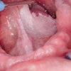
 In stage T3, carcinoma cells invade the muscle layer of the tongue; these cells have a strong positive reaction for the MUC1 glycoprotein. Image courtesy of Rie Kawashima, Jichi Medical University.
In addition, information about the condition of the external environment is communicated to epithelial cells via signal transduction through membrane-associated MUCs.
This means they may serve as cell-surface receptors and sensors that induce cell proliferation, differentiation, and apoptosis, according to lead study author Rie Kawashima, a student in the oral and maxillofacial surgery department at the Jichi Medical University dental school in Japan.
The MUC family comprises large, secreted, gel-forming and transmembrane (TM) mucins, Kawashima explained. Of these TM mucins, MUC1, MUC4, and MUC16 have been shown to be overexpressed in various malignancies.
MUC1 has long been viewed as a tumor-associated molecule because of its frequent overexpression and glycosylation in most carcinomas. Indeed, MUC1 is overexpressed in more than 90% of breast cancer cases and frequently in ovarian, lung, colon, and pancreatic cancer, the researchers noted.
MUC1 is associated with invasive growth of various types of cancers and poor outcomes for patients, they added.
"There is a strong correlation between the expression of MUC1 and patient survival," Kawashima said. "It is considered to be a useful prognostic factor for poor outcomes in a variety of cancers."
Immunohistochemical staining
For this study, the researchers used immunohistochemical staining of MUC1 to analyze oral biopsy specimens from 30 patients (13 men, 17 women) with tongue cancer who were diagnosed and treated at the Jichi Medical University Hospital. The average patient age was 60.5 years.
The degree of staining for MUC1 in tumor cells was evaluated by one oral pathologist who was not aware of the background clinical data. The cases were classified into four groups based upon the degree of staining (0, negative staining of tumor cells; +1, weak expression; +2, moderate expression; +3, strong expression). Tumor status was evaluated according to the World Health Organization's TNM classification system (T0-T4).
Based upon the pathologist's analysis, the overall expressive of MUC1 in the biopsy specimens was 60%, the researchers reported. MUC1 expression was observed mainly at the cell membrane but also in the cytoplasm of cancer cells. The percentage of MUC1-positive specimens in the group with larger tumors (T3 and T4) was much higher (86%) than in the group with smaller tumors (T1 and T2) (52%).
In addition, a higher percentage of MUC1 expression was shown in the advanced mode of invasion (4C) group (71%) compared with that of the mode of invasion (2+3) group (56%). The percentage of MUC1-positive specimens in the postoperative neck lymph node metastasis group (75%) was higher than that in the group with no metastases (54%). However, it was not statistically significant, researchers noted.
"MUC1 overexpression may be associated with the invasive or metastatic properties of cancer cells, resulting in a poor prognosis for patients with MUC1 expression," the researchers concluded. "MUC1 was preferentially expressed in advanced and metastatic squamous cell carcinoma of the tongue and appears to be a predictive marker for lymph node metastasis."


