Tomosynthesis -- a technique for producing slice images using conventional digital x-ray systems -- is not a new concept, but combining it with a dental panoramic imaging system to improve image quality regardless of patient positioning is.
|
A type of tomography Digital tomosynthesis is an old engineering idea that gained new life with the advent of flat-panel digital radiography detectors in the 1990s. It is a form of tomography that enables imaging data from a 2D x-ray to be processed into "slices" that show anatomic structures at different depths and angles. Because the image processing is digital, the slices can be reconstructed from a single image acquisition, saving time and reducing radiation exposure. In recent years, most of the tomosynthesis research has focused on using the technology for early detection of breast and lung cancer. |
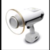
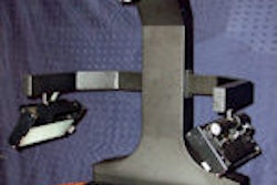
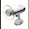
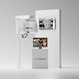
Researchers at the University of Texas Health Science Center (UTHSC) at San Antonio, Hosei University, and Showa University have been working with the PanoACT-1000 panoramic system, which incorporates a PC-1000 unit (Panoramic) with a cadmium telluride (CdTe) detector and a digital signal-processing technique based on tomosynthesis (Dentomaxillofacial Radiology, January 2010, Vol. 39:1, pp. 47-53). Their goal is to develop a practical method of reconstructing high-quality panoramic images without having to worry about patient positioning; in initial experiments at UTHSC, the system allowed the extraction of an optimum-quality panoramic image regardless of irregularities in patient positioning, the researchers wrote.
"Say a patient is positioned in the panoramic machine, but they end up being too far forward or too far back," said Robert Langlais, D.D.S., M.S., a professor at UTHSC and co-author of the Dentomaxillofacial Radiology article. "The image will be corrupted because the patient was properly positioned. With tomosynthesis, and using a CdTe sensor, the PanoACT system has a much faster image capture rate compared to film, phosphor plates (PSPs), or CCD sensors. The rest of it is up to the software."
Autocorrection and radiation reduction
And what the software is designed to do is unique, Dr. Langlais said.
   |
| Click here to enlarge these images. |
|
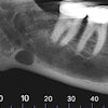
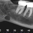
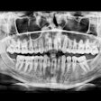
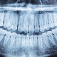
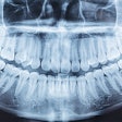
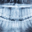
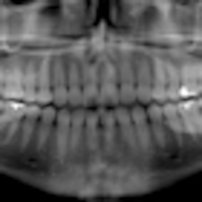
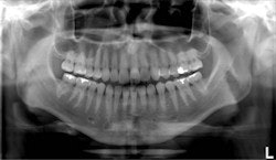
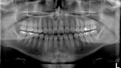
Top, standard panoramic image from a PC-1000 system using a CdTe sensor from Ajat and the PanoACT-1000 tomosynthesis software. Middle, although the anterior teeth are distorted, the posterior teeth no longer have overlapped interproximals. By manually positioning the patient in the system, the contacts are now open. Bottom, the tomosynthesis software then autocorrected the image to bring the front teeth into focus. All images courtesy of Dr. Robert Langlais. |
"In ordinary digital imaging, the software can make the image lighter or darker, change the contrast, magnify, zoom, copy, etc.," he said. "This software goes beyond that. It will autocorrect for positioning errors and generate a new image that is better than the original image. This is a software function that other digital imaging software just can't do."
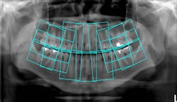
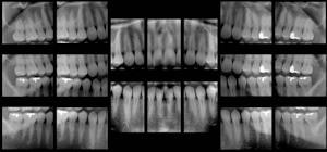
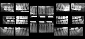
In addition, the software can take a single panoramic x-ray and automatically divide it up to display as a full-mouth intraoral survey, extracting 18-20 regions of interest from a single pano image. This opens up the possibility of using this technique for posterior interproximal caries detection, he added.
"Everyone in the dental profession believes that the bitewings are the gold standard for finding cavities between the back teeth," Dr. Langlais said. "Panoramic images overlap the contact points and don't provide as much detail as the intraoral bitewing images. But in tomosynthesis, we've experimented with getting the patient into a position where the interproximal contacts are open. We have been successful in taking a [pano] machine and acquiring the whole scan with the contacts open."
 |
| Click here to enlarge this image. |
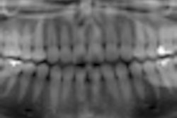
| The software also identifies regions of interest where 18 intraoral images would normally be taken, then extracts the images from a single panoramic x-ray to create an 18-image survey. |
He and his colleagues plan to publish a thesis on this that shows no significant difference between the panoramic bitewings and ordinary bitewings, he added. Such an advance could eliminate the need for intraoral bitewings, which can be uncomfortable, time-consuming, and overexpose the patient to radiation.
"A single digital pano image is five to 10 times less radiation than digital intraorals," Dr. Langlais said. "So, conservatively, the image of the pano system with tomosynthesis is five times less radiation than intraorals."
Even so, he is in the minority when it comes to his ideas about using this technology to make it possible for panoramic imaging to replace intraoral bitewings.
  Click here to enlarge these images. |
"Dr. Langlais, a highly respected oral and maxillofacial radiologist, is interested in something we are all interested in: finding a way of detecting caries without having to place something in someone's mouth," said Don Tyndall, D.D.S., M.S.P.H., Ph.D., director of oral and maxillofacial radiology at the University of North Carolina at Chapel Hill School of Dentistry, in an interview with DrBicuspid.com. "Dr. Langlais has stated in his textbook on panoramic radiology that panoramic imaging is equal to intraoral bitewing images for detecting caries. Unfortunately, at this time there is no published research that scientifically validates this claim. So while I applaud the idea of extraoral caries detection, and it is worth pursuing, I don't think there is any scientific evidence yet to show there is anything better at this point than intraoral radiography."
However, Dr. Tyndall added, "I do believe panoramic-based bitewings are a step in the right direction."
Copyright © 2010 DrBicuspid.com
