Cone-beam CT (CBCT) helped identify a rare case of a dental implant moving into a man's right ethmoidal sinus in a recent case report, published online on May 27 in Heliyon. An anatomical variant likely prevented him from showing signs of sinusitis related to the dislocated implant.
The variant of a bilateral accessory maxillary ostium may have provided an additional way for mucus to drain in the maxillary sinus, allowing the implant to move into the ethmoidal sinus without causing the common symptom of mucus stagnation, which leads to sinusitis. Taking CT scans after implant surgeries may prevent this, according to the authors.
"We ... suggest performing a 3D CT scan in all cases after a dental implantation, in order to disclose the correct placement of the implant and to look for anatomic variants of the sinus in slightly symptomatic patients," wrote the group, led by Filippo Cascio, MD, of the department of otorhinolaryngology at Papardo Hospital in Messina, Italy.
Unusual but not impossible
Anatomical variants of patients' paranasal sinuses can lead them to develop rhinosinusitis. Though it is unclear why, the accessory maxillary ostium is a variant found in patients with chronic rhinosinusitis. Dental implants and endodontic materials impinging on the maxillary sinus can cause maxillary sinusitis, though it is rare. Maxillary sinusitis may result in headaches that often occur near the sinus, a foul-smelling nasal or throat discharge, and fever or weakness.
There have been rare cases in which endodontic materials were entirely displaced into the maxillary sinus, which led to sinusitis due to an obstruction of the maxillary ostium. Until now, the effects of paranasal sinus variations on dental implant dislocation have not been described, the authors wrote.
A 63-year-old man
The patient went to the hospital because he was experiencing a feeling of heaviness in his right maxillary sinus and had trouble recognizing smells. The symptoms started after he had an osseointegrated dental implant placed in his 1.5 upper right molar, which occurred 20 days prior to his visit to the hospital. He didn't have any symptoms that suggested he had implant dislocation related to maxillary sinusitis, according to the authors.
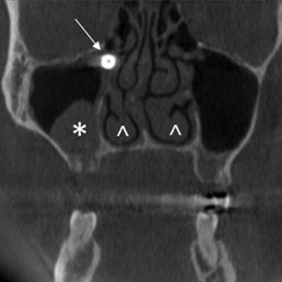
Based on a clinical exam, he did not appear to have acute sinusitis, nasal polyps, purulent secretion coming from the middle meatus, or oroantral communication. Because his symptoms were disabling and worsening, he underwent a 3D CBCT scan. It showed that a 10 x 15-mm dental implant was displaced in the right ethmoidal infundibulum of his right maxillary sinus. It was associated with a mucus retention cyst in the right maxillary sinus and mucosal hyperplasia in the floor of the left maxillary sinus, the authors noted.
 CBCT images in coronal (A), axial (B), and sagittal (C) sections show the implant located at the ethmoidal infundibulum level of the right maxillary sinus. The asterisk shows a mucus retention cyst in the right maxillary sinus. The arrowheads show the asymmetry of inferior turbinates, which may be due to compensatory hypertrophy after the right septal deviation. Image courtesy of Filippo Cascio, MD, et al. Licensed under CC BY-NC 4.0.
The man was placed under general anesthesia and the implant was removed, they wrote. He reported no other symptoms after three days.
Clinicians should consider that relatively common anatomical variants of the paranasal sinuses, such as an accessory maxillary ostium, could alter the underlying physical processes associated with disease and lead to unusual clinical presentations, according to the authors.
"We would suggest considering a possible dental implant dislocation even without clear rhinosinusitis symptoms, since there could be anatomic variants that may avoid the collection of mucus in the sinus," they wrote.

