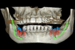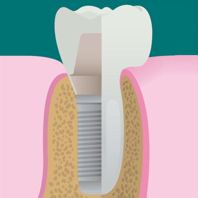
Successful implant treatment requires high-resolution images at all three treatment phases: planning, surgery, and follow-up. As ultrasound is a promising but not widely studied imaging modality for dental implants, researchers wanted to see if it had practical application
Ultrasound can be a potentially useful clinical tool in all three treatment phases, they found, but the team also cautioned that more study and guidelines are needed, especially in the area of implant stability imaging.
"Interested stakeholders, e.g., researchers, dentists, ultrasound manufacturers, and entrepreneurs are encouraged to collaborate to bring ultrasound into clinics for the welfare of millions of patients," the authors wrote (Dentomaxillofacial Radiology, May 23, 2018).
The study was led by Vaishnavi Bhaskar, BDS, MPH, a student in the internationally trained dentist program at the University of Michigan School of Dentistry in Ann Arbor.
Miniature probes
With the increased use of dental implants, the need for high-quality, nonintrusive, and low-cost images of the implant site has increased.
“Interested stakeholders ... are encouraged to collaborate to bring ultrasound into clinics for the welfare of millions of patients.”
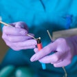
Ultrasound imaging is being used in almost every field of medicine, but challenges such as the difficulty of fitting an ultrasound probe in a patient's mouth and the technology's resolution have prevented it from making inroads into dentistry. However, the introduction of miniature-sized probes with superior image quality may change that.
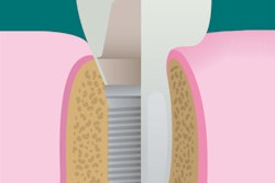
During each treatment phase, accurate knowledge of soft- and hard-tissue dimensions, relationships to vital structures, and bone density measurements are prerequisites for successful implant placement. The researchers of the current study proposed that ultrasound has the potential to emerge as a useful imaging tool in implant therapy, and they wanted to investigate potential indications of its use in the three treatment phases of implant dentistry.
To test their proposal, they conducted a systemic literature review, reducing their initial list of more than 400 articles to a final list of 28.
Based on their literature search, the group found that ultrasound could be potentially useful in all three treatment phases. In the planning phase, the researchers found that the modality could evaluate vital structures in the mouth, tissue biotype, ridge width, ridge density, and cortical bone thickness in and around the implant site.
During surgery, ultrasound can provide feedback by identifying vital structures and bone boundaries at the implant site, the authors reported. At follow-up, it can evaluate marginal bone level and implant stability (see table below). They did caution that research on the modality's use in measuring implant-bone stability is in the early stages.
| Ultrasound indications based on phases of implant dentistry | |
| Treatment phase | Potential indications |
| Planning |
|
| Surgical |
|
| Postoperative follow-up |
|
The authors noted that ultrasound does not image inside bones, so it cannot be used to diagnose hard-tissue pathology, for example. Also, the modality is able to image a focused site in 2D but not the entire jawbone, meaning it is best indicated for single implant restorations.
Future focus
The authors listed no study limitations.
They suggested that future research could focus on evaluating implant wound healing and peri-implantitis activity with ultrasound, but concluded their literature review found potential indications for ultrasound's use with dental implants.
"For the first time, this systematic review successfully identified many potential indications for ultrasound use in implant therapy," the authors wrote.
