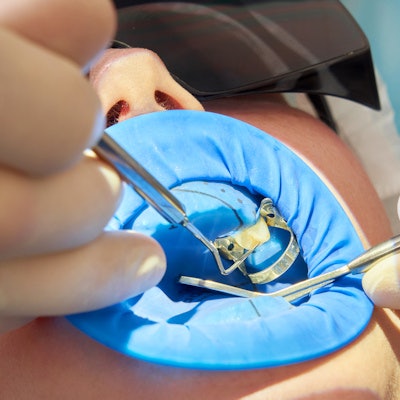
A stainless-steel rubber dam clamp used during a root canal may have led to cortical necrosis in a healthy 22-year-old woman's mandibular tooth, according to a case report published on March 10 in the Journal of Endodontics.
It is believed to be the first published report that shows the effect that a rubber dam clamp may have on underlying bone in healthy patients, the authors of the case report wrote. After a rubber dam clamp is removed following a dental procedure, clinicians should document the position of the tooth isolation brace and evaluate alveolar tissues.
"A rubber dam clamp placed onto gingiva overlying alveolar bone may cause cortical bone necrosis, sequestrum formation and abscess or foreign body reaction in otherwise healthy individuals," wrote the authors, led by Dr. Peter Cervenka, an oral and maxillofacial surgeon at the Naval Dental Center in Norfolk, VA.
22-year-old woman with intense jaw pain
After two weeks of experiencing lower right jaw pain, the woman visited the dentist. Her vital signs were normal, but an x-ray revealed that tooth #31 had symptomatic irreversible pulpitis and periapical periodontitis.
The patient was anesthetized, and a nonlatex rubber dam and stainless-steel clamp were applied to tooth #31. A clinician performed a pulpectomy, irrigated the canals, and informed the patient to take acetaminophen and ibuprofen for pain. Also, she was told to see an endodontist to have the root canal completed, according to the report.
Four days later, she visited an endodontist for treatment. Her tooth again was isolated with a rubber dam and clamp, and the root canal was completed. She was told to return to her dentist for a tooth restoration, the authors wrote.
A week later, she returned to her dentist for the restoration. When she arrived, tooth #31 presented with inflamed lingual gingiva, so the restoration was delayed for four weeks. Nearly three weeks later, the woman went to her dentist with a complaint of three days of low-grade pain intensity and a pressurelike feeling with swelling near tooth #31.
An exam revealed localized swelling with purulent drainage on the midroot region, which was lingual to tooth #31. The patient underwent periapical x-rays, which revealed lucency in the mesial and distal roots of the tooth.
After the tooth was diagnosed as previously treated with a chronic periapical abscess, the patient chose to have it drained and to seek endodontic consultation. She also underwent a cone-beam computed tomography scan (CBCT), the authors wrote.
About three weeks later, she was seen by a second endodontist. At this visit, the woman explained that a small foreign body had dislodged from the site of the tooth, eliminating her pain, and returning her gums to normal. After the endodontist examined her and found no signs of swelling or infection, he referred the patient to her dentist for a restoration.
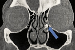
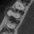
Later, a radiologist interpreted her CBCT, noting that tooth #31 had a normal periapical appearance. However, the woman's "adjacent lingual cortical bone had a lytic and erosive appearance" that stretched about 16 mm in anteroposterior dimension by 7 mm craniocaudally that was limited to the thickness of the lingual cortex.
The radiologist interpreted the findings as possible osteomyelitis of tooth #31 and relayed this information to the patient's general dentist. After consulting with an oral surgeon, the woman was referred to the treating endodontist to review the CBCT, they wrote.
Five days later, the patient returned to the original endodontist, complaining of sensitivity, as well as tenderness and pressure when biting. There was no swelling, but tooth #30 was "responsive but lingering to cold testing." The original endodontist review of the CBCT, as well as that of the radiologist, revealed a hyperdensity near the tip of tooth #30 with an untreated root, distobuccal tooth #31, the authors wrote.
The patient was diagnosed with "symptomatic irreversible pulpitis and condensing osteitis of (tooth) #30 and previously treated with an untreated root, symptomatic apical periodontitis tooth #31," according to the report.
The endodontist diagnosed the woman with an iatrogenic injury from the rubber dam clamp, not osteomyelitis. Furthermore, CBCT images revealed a concave outline of missing lingual cortical bone that matched the contour of the stainless-steel clamp. A root canal was performed on tooth #30 and the untreated root of tooth #31, the authors wrote.
Over the following months, the patient was monitored by her dentist. She made a complete recovery, and at her six-month follow-up visit, a CBCT scan showed normal lingual cortical bone. Finally, full gold crowns were placed on both teeth, they wrote.
An uncommon problem
Clinicians use rubber dams during dental procedures to help reduce microbial contamination and decrease a patient's potential for ingesting or inhaling instrumentation, debris, or chemicals. When using a dam, a clamp often is required. However, the clamp can be a source of complications, including gingival injury and osteonecrosis in susceptible individuals, they wrote.
"This case details changes that can occur in mandibular cortical bone architecture following the use of a stainless steel rubber dam clamp during an endodontic procedure," Cervenka and colleagues wrote.


