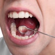
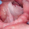
In vivo Raman spectroscopy is showing promise as a diagnostic tool for identifying malignant and potentially malignant lesions of the human oral cavity in a clinical setting, according to a study in the Journal of Biophotonics (July 2, 2013).
The study, conducted at the Raja Ramanna Centre for Advanced Technology in India, involved 28 healthy volunteers and 171 patients having various lesions of the oral cavity.
The researchers measured the Raman spectra from multiple sites of normal oral mucosa and lesions belonging to squamous cell carcinoma (OSCC), oral submucous fibrosis (OSMF), and leukoplakia (OLK). They then subjected the spectra to a probability based multivariate statistical algorithm capable of direct multiclass classification.
With respect to histology as the gold standard, the diagnostic algorithm was found to provide an accuracy of 85%, 89%, 85%, and 82%, respectively, in classifying the oral tissue spectra into the four tissue categories, the study authors reported.
In addition, the algorithm resulted in a sensitivity and specificity of 94% in discriminating normal from the rest of the abnormal spectra of OSCC, OSMF, and OLK tissue sites pooled together.
