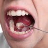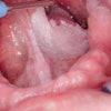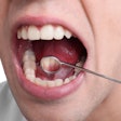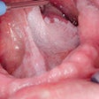
SAN FRANCISCO - While commercially available optical tools can help clinicians identify premalignant lesions in the oral cavity, the prevalence of confounding lesions increases the rate of false positives, according to researchers at this week's SPIE BiOS meeting.
"Compared to any other body site, the oral cavity has the highest number of confounding lesions," said Nadarajah Vigneswaran, BDS, DMD, a professor of diagnostic sciences at the University of Texas Dental Branch at Houston.
With 300,000 new cases of oral squamous cell carcinoma (OSCC) occurring annually worldwide, diagnosis at an early stage is critical to improving survival, Dr. Vigneswaran noted. However, screening via visual examination and palpation by general dentists during routine dental exams has resulted in poor detection rates. In addition, dentists frequently detect white or red patches during routine screening of an asymptomatic patient that turn out not to be premalignant lesions, according to Dr. Vigneswaran.
“Loss of autofluorescence can occur in confounding lesions just as in premalignant lesions.”
— Nadarajah Vigneswaran, BDS, DMD,
University of Texas Dental Branch
at Houston
"Most OSCC is preceded by clinically evident potentially malignant lesions," he said -- notably leukoplakia, which presents as white patches, and erythroplakia, which presents as red patches. "But dental practitioners do not have the clinical training and experience to distinguish potentially malignant lesions from confounding lesions, nor do they have tools for differentiating between premalignant lesions and nonpremalignant lesions."
Many noncancerous conditions can present as white or red patches, Dr. Vigneswaran explained. For example, most red lesions are inflammatory in nature and can represent -- in addition to erythroplasia -- candidiasis, avitaminosis, angiomas, burns, purpura, lichen planus, and telangiectases (British Medical Journal, July 22, 2000, Vol. 321:7255, pp. 225-228). Most white lesions are keratoses caused by cheek biting, friction, or tobacco use, although other conditions can include infections (such as candidiasis, syphilis, and hairy leukoplakia), dermatoses (usually lichen planus), and neoplastic disorders (such as leukoplakias and carcinomas).
Oral cancer and precancer display a loss of autofluorescence across a broad range of ultraviolet and visible excitation wavelengths, and autofluorescence has been shown to enhance the assessment of these lesions, Dr. Vigneswaran noted. However, while autofluorescence detection devices such as the Velscope/Velscope 3x, Identafi 3000, and DOE (dental oral examination system) can help dentists identify premalignant lesions without having to biopsy every suspicious lesion, clinicians need to familiarize themselves with the confounding lesions that contribute to the high false-positive rates seen with these devices.
"Loss of autofluorescence can occur in confounding lesions just as in premalignant lesions, which is one of the diagnostic challenges," he said.
Next-generation devices that can help differentiate between true premalignant lesions and mimicking lesions "would be very valuable for clinicians," Dr. Vigneswaran said.
"Developing and validating an acceptable noninvasive diagnostic test that can discriminate benign oral mucosal lesions from oral cancers and its precursors with minimal false-positive and false-negative results would be beneficial not only for the patient but also to society by reducing healthcare costs through avoiding unnecessary scalpel biopsies," he and colleagues from Rice University and MD Anderson Cancer Center noted in a 2010 study in Future Oncology (July 2010, Vol. 6:7, pp.1143-1154).
Toward that end, they are investigating multimodality approaches to enhance the early detection of oral cancer and reduce the false-positive rates, combining high-resolution fluorescence imaging with wide-field imaging to improve specificity by imaging subcellular detail of neoplastic tissues.
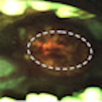
Tissue autofluorescence of common oral mucosal lesions
|
Copyright © 2011 DrBicuspid.com
