A 29-year-old healthy man experienced a rare case of losing six teeth followed by upper jaw osteonecrosis due to an infection with shingles, a rash caused by the same virus that causes chickenpox. The case report was published on July 27 in BMC Oral Health.
The case highlights the importance of healthcare professionals, specifically dentists, knowing the precursor signs of the herpes zoster (HZ) infection, commonly referred to as shingles, to avoid needed procedures or delays in treatment, the authors wrote.
"HZ infection of the face should be taken very seriously to obtain prompt treatment to prevent the rare complications of bone necrosis and tooth loss as much as possible," wrote the authors, led by Jianfeng He of the Zhejiang University School of Medicine in China.
A 29-year-old man who lost an upper incisor
In March 2021, a healthy man with no history of chronic diseases visited an outpatient clinic after losing his central upper incisor about 12 hours earlier. He had clusters of erythema and papules, along with blisters, that arose from a small blister on the left side of his face and upper palate, about two weeks before he lost the tooth. He reported a burning sensation on the side of his face and upper palate, which progressed into painful ulcers. He had no significant medical history other than having chickenpox during childhood, the authors wrote.
 (A) An extraoral photograph (frontal view) showed multiple irregular, crusted lesions on the left face extending along the rami maxillary trigeminal nerve. (B) An intraoral photograph of the empty extraction fossa with smooth inherent alveolar bone, devoid of bleeding, and several large ulcerative surfaces in the mucosa. (C) An intraoral photograph captured swelling, congestion, and erosion in the upper left hard palate. Images and captions courtesy of Sun and colleagues. Licensed under CC BY 4.0.
The man had irregular superficial sores and scabs on his lip, nostrils, nasal wing, and suborbital region. Also, he had other ulcers of varying sizes on his lip and mucosa, as well as severe erythema, edema, and congestion on his left upper gum.
In the area where his incisor fell out, exposed alveolar bone in the socket without any signs of bleeding or pus was visible. Imaging revealed no necrotic bone in the cavity in the upper left jaw.
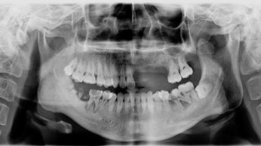 A panoramic radiograph taken during the man's first visit.
Based on the patient's medical history and clinical data, a preliminary diagnosis indicating herpes zoster of the face with involvement of the maxillary branches of the trigeminal nerve was made. He was prescribed famciclovir, gabapentin, and metronidazole, the authors wrote.
About one month after his initial tooth loss, dead bone was detected in the edentulous area, and it was completely resected. Finally, the man was diagnosed with osteonecrosis caused by shingles, they wrote.
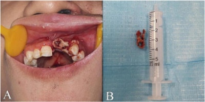 (A) An intraoral photograph showed the presence of sequestrum formation in the area of the left upper anterior teeth. (B) The removed sequestrum.
Four months later, he lost his second premolar. His canine, first premolar, and third molar were very loose, so they had to be extracted.
Approximately one year after his diagnosis, his facial scars and pigmentation had subsided, and his damaged mucosa had recovered. However, an exam revealed there was still distal alveolar bone loss around the second upper left molar, the authors wrote.
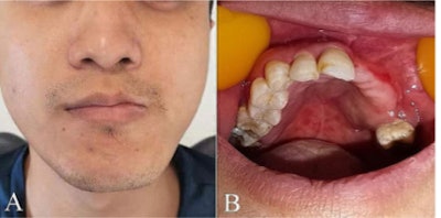 (A) An extraoral photograph showed the facial scars and hyperpigmentation had mostly subsided. (B) An intraoral photograph showed the full recovery of mucous membranes and gums.
An unusual complication
HZ, a virus that occurs with varicella-zoster virus reactivation, has an incidence rate of 1 in 1,000 young people, however, the occurrence increases up to tenfold in the elderly. Though the viral infection can cause complications, including burning pain in the nerves and skin, it is not linked typically with osteonecrosis or tooth loss, according to the report.
"Education regarding the uncommon complications associated with trigeminal herpes zoster infection remains necessary," He and colleagues wrote.


