One of the promises of digital dentistry for implants is that the process is faster and more accurate. But the results of a new study might temporarily alter that statement.
Which is more accurate for implant placement: a virtual model reproduced by an intraoral scanner or a conventional silicone impression? Researchers from Japan undertook the first study that measured the accuracy of an optical impression scanner compared with conventional impressions for this placement.
"In the near future, the development of information technology could enable improvement in the accuracy of the optical impression with intraoral scanners," the authors wrote (PLOS One, October 5, 2016).
Scanner versus impressions
While many published reports have described the accuracy of using the optical impression technique to fit crowns or bridges, the researchers of the current study wanted to see if using an optical scanner was as accurate at implant placement as models cast by conventional silicone impression, a technique familiar to many dentists. An optical intraoral impression system can reproduce shapes of cavities, abutment teeth, and adjacent teeth, according to the authors.
The researchers placed two implants (Nobel Biocare) on a master model: one on the second premolar and the other in the first-molar region. The abutments were 5 mm and 7 mm in height. Working plaster casts were made from the master model by silicone impression.
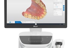
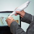
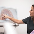
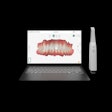
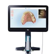
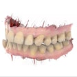
The model was also scanned using an intraoral scanner (Lava Chairside Oral Scanner C.O.S., version 3.0.2, 3M ESPE). The accuracy was measured using a computer numerical control coordinate measuring machine as a control. The master model was scanned 10 times with the scanner and also 10 times with the measuring machine.
The researchers measured the distance between two ball abutments that were connected to the implants. They found that the "trueness of distance error" between them was 64.5 µm for the scanner and 22.5 µm for the working casts, which means the error of working cast was smaller than that of the scanner. The range of the distance error was 27.7 µm to 92.2 µm for the scanner and 1.7 µm to 40.8 µm for the working casts using the measuring machine.
“The development of information technology could enable improvement in the accuracy of the optical impression with intraoral scanners.”
The range of distance error for the working casts and scanner was similar (scanner from 1.5 µm to 36.9 µm; model from 2.0 µm to 32.0 µm), the authors noted. There was no significant differences for precision between the scanner and cast, they concluded.
The researchers also determined the angulation error for both the 5- and 7-mm-height abutments. They found that the mean angulation error of the scanner was greater than the error of the working cast at the 5-mm height, which they concluded suggested that "trueness and precision of the optical impression is inferior to conventional impression."
The same was not true, however, for the 7-mm-height healing abutments, as there was no significant difference in the angulation error of the scanner from the working cast in trueness and precision. The authors suggested that "the height of healing abutment may affect the error level for angulation in the Lava C.O.S."
Limited phenomenon?
The authors noted that a 2013 study found that digital impressions were "not as true or precise as conventional impressions." The authors of that study suggested that, right now for restorative procedures, digital impressions could not replace conventional impressions.
However, the results from the current study may be only a "limited and temporal phenomenon because the trends in digital dentistry are drastic," the authors noted. They suggested that further analysis is needed because of the new devices entering the market that should improve measurement accuracy.
"In this study, distance error of the optical impression was slightly greater than that of conventional method," the authors wrote. "Using longer healing abutment, the trueness and precision of angulation error were improved in the optical impression."
