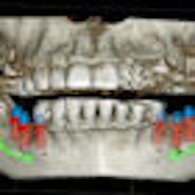
As increasingly sophisticated imaging technologies make their way into dentistry, the likelihood of incidental findings showing up on a 2D or 3D study increases as well -- along with potential medicolegal issues for practitioners who miss them, according to a systematic review in the current Journal of the American Dental Association (February 2013, Vol. 144:2, pp. 161-170).
"It remains the responsibility of the clinician to analyze the entire volume of data, and not just the region of interest, to avoid missing a significant finding regardless of the imaging modality used or the image size generated," wrote the study authors, from the University of Alberta.
Quantifying the frequency
To determine the frequency and nature of incidental findings in the head and neck region on cone-beam CT scans, the researchers systematically searched Medline, Embase, PubMed, Scopus, Web of Science, and the Cochrane Library databases through mid-July 2012.
"Quantifying the frequency of incidental findings in 3D radiography may affect evolving cone-beam CT guidelines and has significant considerations for both the doctor, in medicolegal terms, and the patient, in terms of the potential diagnosis of as-yet-undetected disease," the study authors wrote.
They found a total of five studies that met their inclusion criteria; all five were written in English and published between 2007 and 2012. After reviewing these studies, they determined that the frequency of incidental findings ranged from 1.3 to 2.9 per cone-beam CT scan or from 24.6% to 93.4% of scans, depending on the reporting method.
The authors noted several possible reasons for the inconsistency in the results of the studies:
- The populations were not standardized.
- There was a wide range in participants' mean age (13.0 to 64.7 years).
- The method of reporting: The frequency of incidental findings can be described either as the absolute number of findings detected or as the number of scans containing incidental findings.
- The studies all used a retrospective, cross-sectional design, which means an inherent risk of potential bias.
Even so, the researchers concluded, "incidental findings are frequent in cone-beam CT imaging, and they vary considerably in respect to their frequency and nature." Based on their literature review, the most common are extragnathic findings, including vertebral degenerative changes, sinusitis, pineal gland calcification, impacted third molars, mucous retention cysts, temporomandibular joint condylar degenerative changes, and concha bullosa.
More research needed
More research is needed to determine the effects of these incidental findings on the need for follow-up care and intervention, as well as the potential cost of additional treatment, they added.
"From this review, we conclude that further investigation must occur regarding the establishment of a standardized method of reporting and definition of the term 'incidental finding' to be used both clinically and in research," the study authors concluded.
“Ethics demand maximizing reading the whole image volume.”
Allan Farman, BDS, MBA, PhD, DSc, a professor of radiology and imaging science at the University of Louisville in Kentucky and immediate past president of the American Academy of Oral and Maxillofacial Radiology, agreed with many of the authors' conclusions.
"I fully concur that there is a need for more extensive studies, perhaps using a multisite approach of datasets where there are thousands or tens of thousands of CBCT images or reports investigated for the incidence of pathoses that significantly affect treatment and patient health prognosis," he stated in an email to DrBicuspid.com.
"Research needs to concentrate on clinically significant findings and the effects of such matters as field-of-view, exposure parameters, and the medical model of overreads on achieving the best patient outcomes," Dr. Farman added. "Ground truth is rarely achieved by radiography alone, so clinical features and histopathology should be included in such research. This is primarily an ethical issue and only secondly a medicolegal one. Ethics demand maximizing reading the whole image volume, and having that appropriately documented is a way to help alleviate legal risk."
Looking ahead, Dr. Farman believes that cone-beam CT will enter the mainstream of dentistry, with all new dental graduates being well-trained in 3D anatomy and basic conditions and also familiar with which cases to refer.
"Likely there will also be a growth in availability of feature-detection software to help via computer-assisted diagnosis," he stated. "For the present, most users did not get this type of training before purchasing and using a dental cone-beam CT unit, so continuing education is especially important, and care is needed in selecting the source of such training."


