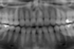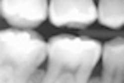
Just as Mark Twain commented about his reported death when reading his own obituary, news of the demise of the dental bitewing radiograph for detecting proximal surface dental caries is a bit premature.
Colleagues have asked us about the report in DrBicuspid.com concerning a study published last month in Caries Research, "Radiographic and Laser Fluorescence Methods Have No Benefits for Detecting Caries in Primary Teeth" (August 16, 2012).
The title of the study, reflecting the authors' results, is diametrically opposite to previously published literature supporting the use of bitewing radiography in children for proximal caries detection: Australian Dental Journal, March 2009, Vol. 54:1, pp. 23-30; European Journal of Paediatric Dentistry, March 2003, Vol. 4:1, pp. 40-48; Pediatric Dentistry, 2008-2009, Vol. 30:7(suppl), pp. 236-237; Guideline on Prescribing Dental Radiographs for Infants, Children, Adolescents, and Persons with Special Health Care Needs, American Academy on Pediatric Dentistry.
In fact, to some, the findings of the Caries Research study may seem like heresy. However, many of the study's results also support previous research. For example, the authors found that the prevalence rate of proximal caries that cannot be detected by visual-tactile examination alone, or "hidden" proximal caries (found by the difference in sensitivity values between the visual-tactile and radiographic techniques) was 33.3%. This suggests that a significant number of proximal carious lesions in the primary dentition will be missed if bitewing radiographs are not exposed.
Accuracy vs. sensitivity and specificity
However, this is only part of the story. The difference and controversy in this study results from considerations of the prevalence of proximal dental caries.
The calculation of sensitivity and specificity as indices of the efficacy of a diagnostic test do not depend on the disease prevalence. Accuracy, however, does. Prevalence is the number of occurrences of a disease (in this case, positive proximal caries lesions) to the number of surfaces at risk in the sample under investigation. Low prevalence reduces the relative contribution of correct positive diagnoses to the accuracy index and increases the contribution of correct negative diagnoses.
The prevalence of proximal carious lesions using a restorative threshold in the current study was 4.2%. Numerous other studies would suggest that the prevalence rate for this sample is extremely low and most likely reflects the cavitation/noncavitation criteria for presence. Overall prevalence has been reported as high as 86% and up to 46% for cavitated lesions (Pediatric Dentistry, January-February 2005, Vol. 27:1, pp. 54-60). In addition, noncavitated carious lesions are much more prevalent than cavitated carious lesions in children (Community Dentistry and Oral Epidemiology, October 1992, Vol. 20:5, pp. 250-255).
If considered purely from a restorative point of view and taken indiscriminately at an early age, as described in the Caries Research study (mean age 7.4 years ± 1.6), bitewing radiography may indeed provide poor overall accuracy. But this is less than half of the story. Bitewing radiography on the deciduous dentition provides additional clinical information not quantified or considered by the authors in this case.
For example, identifying incipient lesions is perhaps just as important in this age group as those lesions that need treatment. Because of the relative thinness of the enamel, incipient lesions involve dentine more rapidly and are just as important to identify as those that result in frank cavitation. In addition, these lesions represent the potential of future dental caries and should not be excluded.
Also, bitewing radiography provides clinicians with patterns of caries activity relative to a specific individual and so are important in the overall assessment of caries activity/risk, predicting future carious activity (International Journal of Paediatric Dentistry, May 2006, Vol. 16:3, pp. 152-160) and in applying radiographic selection criteria. Exclusion of incipient lesions from consideration and lack of description of dental caries pattern greatly reduce the external validity of the results of the study.
Viewing conditions critical
No study is perfect. However, the Caries Research study also does not provide intra- and interexaminer reliability indices, does not establish the power of the study (Caries Research, May-June 2003, Vol. 37:3, pp. 200-205), and uses a soon-to-be outmoded image capture technique (film).
In addition, apparently the observers did not use magnification when viewing the radiographs. Viewing conditions -- including magnification, luminosity of transmitted light, masking of extraneous light, and ambient lighting -- have substantial impact on the ability to detect dental caries, as do technical quality of image receptor positioning, exposure parameters, and processing quality assurance. In addition, alternative detectors (solid-state or photostimulable phosphors) could permit image enhancement, and computed guidance could be provided.
As a result, this relatively simplistic study cannot be generalized to modern x-ray detection modalities used for intraoral radiography.
The basis for judging a technique has to be using that technique optimally and with adequately trained observers. Dental caries lesions not being detected in a bright clinic on radiographs when not optimally displayed can often be found in a room with reduced ambient lighting and image adjustment or computer-assisted diagnosis algorithms.
Unfortunately, the publication of this type of manuscript muddies the evidence-based pond and potentially does more harm than good.
William Scarfe, DDS, is a professor of radiology and imaging science at the University of Louisville School of Dentistry. Allan Farman, BDS, MBA, PhD, DSc, is a professor of radiology and imaging science at the University of Louisville in Kentucky.


