DrBicuspid.com is pleased to present the next installment of Leaders in Dentistry, a series of interviews with researchers, practitioners, and opinion leaders who are influencing the practice of dentistry.
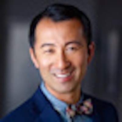
We spoke with Ernest Lam, DMD, PhD, a professor of oral and maxillofacial radiology (OMR) at the University of Toronto, who obtained his DMD from the University of British Columbia in 1989 and then went on to get his specialty training in oral and maxillofacial radiology and a PhD in radiation biology at the University of Iowa.
In 1998, Dr. Lam returned to Canada to take a teaching position at the University of Alberta, where he stayed until 2005 before accepting his current position at the University of Toronto.
Dr. Lam publishes and lectures regularly on critical issues in oral and maxillofacial radiology and radiation biology, such as the role of cone-beam CT in dentistry and radiation dose. He has also become a vocal proponent of the need for more OMRs to go into academics at a time when many are opting instead for private practice.
DrBicuspid.com: What prompted you to become an OMR and go into teaching?
Dr. Lam: One of the reasons was that when I was a dental student, I was a bit indecisive about what I wanted to do in my life. My undergraduate degree was in chemistry, and one option was to become researcher. In my third and fourth years as an undergrad I became very interested in nuclear magnetic resonance (NMR) imaging. I heard about people using this technique to image humans, and I thought, "How cool is that?"
But I ended up applying to dental school because I thought it would be the responsible thing to do. While there, I met a professor who was doing imaging of the jaw muscles as part of his research, and he kind of hooked me into doing my master's degree with him part-time while I was going to dental school. My project was NMR and spectroscopic imaging of jaw muscles. I hated dental school, so doing this research in his lab was what kept me going all those years.
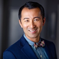 Ernest Lam, DMD, PhD.
When I finished my DMD and master's, I told my boss I was interested in going into academics and he told me I had to get into a specialty and get my PhD. With my background, I thought 3D imaging was going to important, and at the time -- this was in 1990 -- the University of Iowa was one of only three or four programs that included 3D imaging in its syllabus.
I think my mother by this time was wondering why I was still in school. I was 35 when I finished my PhD, and by then everyone else I knew was in practice. That was the model my parents knew, and I think they were quite concerned for many years.
When I finally finished school, I decided to go back to Canada. I got a great education in Canadian schools and thought it was important to come back and contribute to the system. So in 1998 I took a position at the University of Alberta, the most northern dental school in Canada, and the coldest. But it was a really good experience. The dental school is quite small -- they only take 32 students into their first-year program, and you can do a lot of things with 32 that you couldn't do with 90. The school was very supportive and gave me a lot of leeway to develop a program for the students.
Why did you hate dental school?
In my era, people in dental school would say, "I don't know why you can't do this because it works in my hands." They would give you these very nonspecific comments, and there was no such thing as evidence-based dentistry in the 1980s -- or if there was, it was a very small part of what was taught or discussed. The general thinking was, "If this is the way I do it, this is the way you should do it." And coming out of a science degree, my feeling was hey, you should have some kind of rationale for this. I have always thought that if you are doing anything with a foundation in science, you need to utilize principles to do what you do.
How has the OMR specialty changed over the past 20 years?
Twenty years ago, most oral radiologists were academics. It is primarily an academic specialty, and my predecessors all had strong commitments to academia. The major focus was teaching and research. Nowadays, with cone-beam CT, it has really changed. Now students who enter our oral radiology program have more opportunities to go into private practice. This puts the specialty on par with any other specialty, which is a huge step forward. Lots of people say to me that mine isn't really a clinical specialty, but truth is, it is. It contributes to the clinical care of the patients.
So with cone-beam CT, a lot of graduates now can say, "I'm going to open up an office and have patient referrals like any other dental specialist office, and I have this equipment that is pretty unique and I have this other specialty skill set." And the opportunity to have this type of clinical practice is really appealing to a lot of people. So we are seeing a lot of students saying to us, as they are graduating, that they are thinking of opening up a private practice. And this is not unique to Canada -- it is also happening in the U.S. and other parts of the world.
That said, what role do you think cone-beam CT should play in dentistry?
I think people are overusing it and it is being overmarketed, although there certainly are defined roles for it, and a number of people have written on what the defined roles are. The European consortium (SEDENTEXCT) has written evidence-based guidelines based on published literature, and the American Academy of Oral and Maxillofacial Radiology has been putting out evidence-based position papers on the use of different imaging modalities for different diagnostic tasks in dentistry, and having an evidence-based body of work for the use of this technology is really great. But it takes time for this to happen -- in the past four to five years, this body of evidence has started to grow substantially, and there are other areas where many different groups have said hey, it is not useful for this and shouldn't be used for this. My greatest concern is that this type of literature isn't as pervasive in the dental community.
The vendors have a piece of equipment, and they have to sell it to make money. That is their primary purpose, and I appreciate that. So at times, I feel that their primary purpose does not align with mine.
There has been much discussion about radiation dose of late. What is your position on this issue?
I know that when we teach radiation biology and radiation protection at dental school, it is typically in the first or second year. So by the time students reach their third year, they've forgotten most of it. Even dentists in practice, many times they only remember the basics. So I worry about that, in particular for children and adolescents because in our population they are the most radiosensitive people. When you image them, particular care has to be given to ensure that whatever approach you choose, you are choosing a technique to keep radiation doses as low as you can. And the basic way to do that is to limit the number of exams you do.
That is the thing I push the most. As we get older, our tissues become less radiosensitive because there are fewer developing cells in our bodies. But children and adolescents are often not in a position to say, "Do you really think I need this?" They don't have the knowledge to be their own advocates, so someone needs to advocate for them. And I am happy to answer those questions when parents ask them. If they understand what the issues are and are willing to have the discussion, I am willing to have the discussion with them.
Do you still read images for dental practitioners?
I do. One of the things I think is very important for any academic is to be part of the practice community where you live. Most full-time academic positions permit you one day of clinical practice per week, and I think most people take that. I did that in Edmonton; there was a radiological practice and I went there one day a week. I still do some reading for them, and for our in-house faculty practice in Toronto. Our in-house faculty practice gets referrals within the school as well as from outside the school, and we have some pretty good equipment here. I am happy with the sorts of things we are able to do here.
Are there new diagnostic technologies/modalities emerging that will find a place in dentistry in the next five to 10 years?
When I was doing my master's, we used to kid and say wouldn't it be great if someone would create a CT for just the head or an MRI for just the head, and now look where CT is. But the MR machines are quite expensive because you are basically buying a giant magnet, and it is critical to maintain that magnetic field and keep it uniformly across the head. So in order for MRI to occur the way CT has in dentistry, cost has to come down to a level that somebody would even think to afford. But there are definitely advantages to MRI -- resolution of soft tissue is most important.
In cone-beam CT, the next big development will be more sensitive sensors so you can see soft tissue. You kind of already can, but you can't resolve them the way you can on a medical CT or MRI. But the technology is already advancing, which is a good thing.
What's the biggest issue facing the OMR profession today?
I think one of the biggest problems this specialty faces is recruiting enough good people into academics. More radiologists are going into private practice, so in spite of our success I see us going down the same road as specialties like orthodontics and periodontics, where no one wants to be an academic at a university. And this is draining on the field. Somebody has to teach and push the research forward.
That is why I do what I do because this is the best way to move the field forward. In fact, when I meet people who don't know me well, and they ask what I do, I never say I'm a dentist or a radiologist -- I say I'm a professor at the university. But if no one is coming down the pike to take over when I (and other academics) decide to retire, I think this is going to impact the specialty negatively. And this isn't the only specialty where this is happening; this is an issue across all of dentistry.


