Orthodontic radiographs appear to promote cell toxicity in the oral mucosa, according to a new study in Dentomaxillofacial Radiology (October 2010, Vol. 39:7, pp. 437-440) -- calling into question once again whether multiple cephalograms are warranted in every orthodontic case.
To better understand the effects of radiographic radiation on the cellular system, researchers from the Methodist University of São Paulo and the Federal University of São Paolo investigated the frequency of micronucleated cells in the oral mucosa of individuals following orthodontic radiography. They also evaluated the cytotoxic effects -- including pyknosis, karyolysis, and karyorrhexis -- in these patients following exposure.
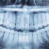
"Orthodontic radiographs carried out in radiological clinics are essential for diagnosis, planning, and control of orthodontic treatment," researchers from Federal University of São Paolo wrote. For example, they noted, both a lateral cephalometric x-ray and panoramic x-ray should be obtained from all patients prior to treatment to enhance the diagnosis and treatment plan.
"Frontal cephalometric analysis is also a valuable complementary examination in establishing the correct diagnosis and orthodontic planning," they wrote. "At present, lateral and frontal cephalograms are considered mandatory in orthodontic therapy."
That said, the possible risk associated with exposure to x-rays must be weighed against the benefits of clinical interpretation, they noted.
"It is well-known that ionizing radiation damages DNA," the researchers wrote. While a variety of assays have been proposed as potential biomarkers, these methods are typically laborious and time-consuming or require highly trained technicians to accurately read and interpret slides, they noted.
So they chose instead to evaluate chromosome damage (micronucleus) and cellular death in exfoliated buccal mucosa cells from patients exposed to cephalometric and panoramic radiographs. Micronucleated cell indexes are thought to reflect genomic instability, and an elevated frequency of micronuclei indicates an increased risk of cancer, they noted.
Small sample
The study comprised 18 healthy volunteers (six male, 12 female) with a mean age of 14.2 years who were referred to the Methodist University of São Paulo for orthodontic therapy. All participants underwent lateral and frontal cephalometric x-rays and panoramic dental radiography prior to orthodontic treatment. Oral mucosa cells were collected immediately before x-ray exposure and 10 days afterward.
"Damage that leads to the formation of micronuclei takes place in the basal layer of the epithelial tissue where cells undergo mitosis," the researchers wrote. "Rapid turnover of epithelial tissues brings the cells to the surface, where they exfoliate. As a result, the maximal rate of micronuclei formation in exfoliated cells is seen one to three weeks after exposure to the genotoxic agent."
Prior to x-ray exposure, the mean frequency of micronucleated cells was 0.02%, the researchers reported. While there was no significant statistical difference (p > 0.05) in the frequency of micronucleated cells following x-ray exposure, an increase in the frequency of karyorrhexis, pyknosis, and karyolysis was observed.
"Despite the lack of cytogenetic damage, our results demonstrated that panoramic and cephalometric radiographs induced cellular death," they wrote. "These results suggest that radiography is able to induce cytotoxicity but not mutagenic effects in oral mucosa cells; therefore, radiographs should be used only when necessary."
Other side effects?
However, while he agrees with the study's overall conclusions, Jay Friedman, D.D.S., M.P.H., author of The Intelligent Consumer's Complete Guide to Dental Health, noted that the researchers did not address some key related issues, including the assumption that all orthodontic cephalograms that are taken will benefit or enhance diagnosis and treatment. He cited a study in Oral Surgery, Oral Medicine, Oral Pathology, Oral Radiology, and Endodontology which concluded that three-quarters of orthodontic radiographs were of no value for either diagnosis or treatment planning (February 1991, Vol. 71:2, pp. 238-245).
In addition, Dr. Freidman said, the current study does not consider the effects of cephalographic radiation on the brain.
"If oral cells are damaged, then is it not likely that brain cells also are damaged?" he said. "We can only conclude that cephalography is not only cytotoxic to oral tissues but also cytotoxic to brain tissues. Therefore, they are contraindicated for routine orthodontic treatment."
Copyright © 2010 DrBicuspid.com