Could an ordinary smartphone be used to identify the oral bacteria that cause dental plaque? University of Washington researchers have converted a smartphone into a mobile multispectral imaging system that lights up plaque through light-emitting diode (LED) images.
The smartphone-enabled system offers a quick, relatively low-cost, easy-to-use method that patients could utilize at home to assess whether they have potentially harmful oral bacteria. The system would be especially useful for those living in rural communities or areas with less access to good dental care, according to Ruikang "Ricky" Wang, PhD, a University of Washington professor of bioengineering and ophthalmology and one of the system's inventors.
"By measuring the size and signal strength of the red-fluorescence area, we believe the patient can be accordingly triaged in terms of its severity to meet different levels of treatments" and aid in tooth decay and disease prevention, Wang told DrBicuspid.com.
 A smartphone's camera was augmented to capture LED-illuminated images. All images courtesy of Ruikang Wang, PhD.
Bacteria assessments are clinically important for oral health because the reproduction of oral pathogens can cause plaque that can lead to tooth decay and periodontal diseases. However, visualizing and identifying the bacteria can be time-consuming and expensive.
Therefore, the researchers set out to develop a new technology that can provide real-time, noninvasive assessment of bacteria. Their findings were published in the May issue of Optics and Lasers in Engineering (Vol. 140, 106546).
An ordinary smartphone
The researchers chose to build their system on a smartphone because of smartphones' widespread use. However, a phone couldn't be used as is. A conventional smartphone contains a camera with red, green, and blue (RGB) channels. An RGB camera funnels different wavelengths of light in the visual spectrum into those three colors.
But bacteria emit many colors beyond blue, green, and red, so they are difficult to view on a conventional smartphone. A black light must be used to get an autofluorescence signal from bacteria, so the researchers knew they needed to get creative, Wang said.
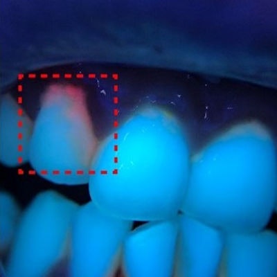
 An RGB autofluorescence image taken with an LED-modified smartphone.
To visualize bacteria, Wang and his team modified the camera of a smartphone by attaching a small, 3D-printed ring that contained 10 LED black lights to the camera opening. Then, they used the augmented device to take images of a person's oral cavity.
When the black light is used, it excites molecules known as porphyrins, which are byproducts of bacterial growth. The molecules emit a red fluorescent signal when they are excited, which allows the pathogens to be visualized, he said.
The LED illumination achieved via the modified smartphone gave the researchers enough data to computationally convert the RGB colors into hyperspectral information, which can be leveraged to see types of bacteria. To demonstrate this method, porphyrin was targeted as the biomarker to identify the bacteria. Converting the information generated a visual spectrum of 15 different sections instead of just red, green, and blue. Typically, expensive, cumbersome lights would be needed to obtain this visual information upfront, Wang said.
 Ruikang "Ricky" Wang, PhD.
"The current ring light provided both the most uniform illumination and appropriate excitation intensity, which is important for quantifiable measurement of the bacteria content in the oral cavity," he said. "While the results were kind of expected, the resulting high imaging quality of the current system is still a surprise to us, simply because of the use of universal and cost-effective smartphones."
For comparison, the researchers used a hyperspectral camera to perform the same task. Compared with conventional multispectral imaging systems, the smartphone-enabled system and method had the ability to perform similar spectral analysis but worked in a snapshot mode, and it was immune to any motion artifacts.
Another benefit is that a "hyperspectral camera is about 30 to 40 times more expensive than the smartphone approach," Wang added.
The researchers plan to further optimize the design and development of the method for commercial use. They also intend to conduct a clinical trial to evaluate its use in the assessment of the bacterial content of dental plaque, he said.
"Ideally, we expect this method to be a selfie screening tool for oral health one day," Wang said.