A woman who experienced tongue pain and taste changes for 18 months felt relief when a rare tumor linked to her lingual nerve was removed. Her symptoms stumped a dentist and were later misdiagnosed as a sialolith, according to a case report published on June 28 in BMJ Case Reports.
Only 6.5% of neural tumors occur in the oral cavity, and there have been very few reported cases of floor-of-mouth solitary neurofibromas, such as in this case. Therefore, clinicians should have a strong understanding of head and neck anatomy before moving forward with diagnoses and treatments, the authors wrote.
This "should help guide the differential for a [floor-of-mouth] mass, which should include lingual neurofibroma," noted the group, led by Alexander Straughan from George Washington University School of Medicine and Health Sciences in Washington, DC.
Solitary neurofibromas are usually benign, and they develop within a major or minor nerve but rarely occur in the oral cavity. When they do develop in the oral cavity, they are usually small and immobile and are found on the lips, tongue, and areas stimulated by the mental nerve. Depending on their location and size, they may cause severe neural pain.
Piercing pain
The 60-year-old woman visited her dentist when she first started experiencing a stinging sensation in her tongue. The dentist could not make a diagnosis, so the patient visited several other specialists. An oral surgeon ordered blood work and other tests, but a diagnosis could not be made. A neurologist ordered a brain scan that revealed nothing remarkable. She was prescribed the nerve pain medication gabapentin, but it didn't help. Meanwhile, her pain, which she compared to having a "red-hot poker" placed through her tongue, grew constant and more severe. She also had trouble tasting food and had a persistent metallic taste in her mouth, according to the authors.
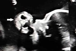
Eventually, she talked to an otolaryngologist about her pain, who referred her to a specialist academic otolaryngology practice. There, she underwent an ultrasound of the submandibular region, which revealed a 5-mm hyperechoic submandibular mass. Unfortunately, these images were not recorded.
Because her Wharton's duct was observed to be dilated proximally with normal vascularity of the submandibular gland, she was given a tentative diagnosis of having a salivary stone and was scheduled to have it removed. A head and neck surgeon attempted to remove the salivary stone but abandoned the procedure once the woman reported sharp neurogenic pain in her tongue despite numbing of her lingual nerve, the authors wrote.
Prior magnetic resonance imaging had failed to identify a definitive cause of her condition, so a computed tomography scan was performed. No definitive mass was revealed, but it did show an asymmetrically prominent possible left facial vein in the submandibular region. The surgeon and patient agreed that an operation should be performed to explore the area and remove the mass.
The woman reported that she felt immediate pain relief after the approximately 4-cm schwannoma was removed from her lingual nerve. Two years later, she has no tongue pain or stinging but experiences some periodic bouts of numbness, Straughan and colleagues wrote.
A teaching moment
When an ultrasound reveals a hyperechoic mass in the floor of the mouth and there is evidence of submandibular duct obstruction, it should not be assumed that the diagnosis is a salivary stone or ductal stenosis. Clinicians should also consider a patient's symptoms. The woman in this case study experienced neuropathic pain, not salivary colic, according to the authors.
"External and lingual nerve-associated masses such as neurofibroma should be considered in differential," they wrote.