Cone-beam CT (CBCT) is considered the gold standard for imaging peri-implant bone, but researchers wondered if another modality without the radiation dose might be more accurate. They tested CBCT against high-frequency ultrasound and reported a surprising conclusion.
Ultrasound had a lower mean measurement error compared with CBCT, while both modalities had the same maximum error, leading the researchers to conclude that high-frequency ultrasound may be useful clinically.
"Within the simulated limited conditions of this study, high-frequency ultrasound, with optical scanning used as a reference, presented higher accuracy in comparison to CBCT, and seems to be a promising tool for measuring peri-implant bone," wrote the authors, led by Juliana Marotti, DDS, PhD, of the department of prosthodontics and biomaterials at the Centre for Implantology at the RWTH Aachen University Medical School in Aachen, Germany (Journal of Clinical Medicine, September 25, 2019).
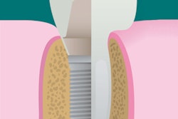
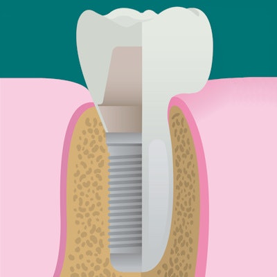
Cone-beam CT is the gold standard for imaging peri-implant bone as it can provide cross-sectional images. However, any artifacts seen on images may jeopardize the viewing of the bone-implant interface and make it challenging to assess peri-implant bone. The researchers also noted that CBCT is not recommended for evaluating asymptomatic implants in periodic exams to avoid patient exposure to high levels of radiation.
Because of these limitations, researchers wanted to see if high-frequency ultrasound could measure bone thickness in the buccolingual region of dental implants with the same accuracy as CBCT. Previous research has shown that ultrasound can reliably evaluate soft tissues, bone surfaces, and dental implants without ionizing radiation, according to the authors.
The researchers placed four dental implants in eight porcine bone samples. Each implant was scanned by a high-frequency ultrasound scanner (a prototype developed at RWTH Aachen University), a CBCT unit (Sirona Galileos, Dentsply Sirona), and an extraoral optical scanner (D250, 3Shape) for comparison.
The table below shows the measurement error obtained from ultrasound and CBCT compared with optical scanning.
| Measurement error for CBCT and ultrasound in 8 bone samples | ||
| CBCT | Ultrasound | |
| Mean value | 0.20 mm | 0.11 mm |
| Maximum value | 0.64 mm | 0.28 mm |
| Highest standard deviation | 0.28 mm | 0.08 mm |
The authors noted that this was an in vitro study and that they chose not to use software to improve image quality as limitations.
The primary significance of the study was to present a new technology for evaluating peri-implant bone, they wrote.
"Ultrasound showed a higher accuracy in comparison with CBCT, while its measurement error was closer to the optical scan values to a small degree," the authors concluded.