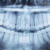
Detecting vertical root fractures in endodontically treated teeth is difficult, no matter the filling material used. A new study investigated whether a tooth's orientation in relation to the x-ray projection plane alters a practitioner's ability to detect these fractures with cone-beam CT (CBCT).
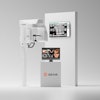
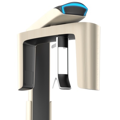
Researchers used a CBCT unit (Picasso Trio, Vatech) and viewed each root with and without different filling materials. They found that the orientation did not matter, irrespective of the filling material used. Their study was published in the Journal of Endodontics (June 1, 2018).
"The detection of [vertical root fractures] using CBCT imaging remains a controversial subject in the scientific literature," the authors wrote. "In general, this diagnosis is more accurate when associating clinical and radiographic signs, and because CBCT imaging provides three-dimensional images, valuable additional information is expected."
The study was led by Victor Aquino Wanderley, DDS, from the division of oral radiology in the department of oral diagnosis at the State University of Campinas Piracicaba Dental School in São Paulo.
Orientation
The detection of vertical root fractures is challenging in the presence of metallic fillings. The accurate diagnosis of these fractures is important to prevent additional damage to periodontal tissues and also to avoid unnecessary treatments and costs.
The researchers wanted to assess the influence of tooth orientation in relation to the projection plane of an x-ray on detecting vertical root fractures with different filling materials using cone-beam CT.
The researchers induced vertical root fractures in 15 endodontically instrumented single-rooted teeth. A control group consisted of 15 teeth similar teeth without vertical root fractures. The teeth were placed into the dental socket of a phantom head. Each root was imaged with the CBCT unit with the longitudinal axis in two orientations: perpendicular and parallel to the x-ray projection plane.
The researchers also scanned each root under three conditions: without filling material, with gutta-percha, and with a metal post.
For the study, five experienced radiologists evaluated the images. The researchers found no significant differences (p ≤ 0.05) in the detection of the fractures, regardless of the filling material. Diagnostic values were similar in both root orientations. The ability to correctly identify those with and without root fracture (Az) did not differ significantly (p > 0.05) between root orientations for any filling condition. It was significantly better (p < 0.05) in the absence of filling material, but it did not differ significantly (p > 0.05) between gutta-percha and metal posts.
| Effect of root orientation and filling material for detecting vertical root fractures on CBCT | ||||
| Root orientation | No filling | Gutta-percha | Metal post | |
| Sensitivity | Perpendicular | 76.0% | 56.0% | 50.0% |
| Parallel | 78.7% | 55.4% | 55.4% | |
| Specificity | Perpendicular | 94.7% | 69.3% | 61.5% |
| Parallel | 89.3% | 59.2% | 50.0% | |
| Accuracy | Perpendicular | 85.3% | 62.7% | 56.0% |
| Parallel | 84.0% | 57.3% | 52.7% | |
| Az | Perpendicular | 0.86 | 0.65* | 0.58* |
| Parallel | 0.86 | 0.62* | 0.57* | |
No material concerns
The authors noted as a limitation that this was an ex vivo study and they did not consider certain clinical and associated radiologic parameters, such as an isolated periodontal pocket and lateral radiolucency. They recommend more studies with multirooted teeth, different endodontic materials, and spatial resolutions.
Nevertheless, tooth orientation for CBCT imaging was not a factor in detecting these fractures, the authors wrote.
"The orientation of the longitudinal axis of the tooth in relation to the projection plane of the x-rays does not influence the detection of [vertical root fractures] in CBCT images irrespective of the intracanal material," they concluded.