Dental x-rays cause a lot more cancer than previously assumed, according to new estimates published in the September Journal of the American Dental Association (September 2008, Vol. 139:9, pp. 1237-1243).
"The dental radiographic procedures we evaluated in this study are 32% to 422% riskier than previously thought," concluded researchers from the University of North Carolina (UNC) at Chapel Hill and the University of California, Los Angeles.
And most dentists aren't doing enough to minimize the risk to their patients, according to lead author John Ludlow, D.D.S., Ph.D., a UNC dentistry professor of diagnostic sciences.
— Allan Farman, B.D.S., M.B.A., Ph.D., D.Sc.
Science has not established a threshold below which radiation is harmless. At the same time, it's hard to show what damage is caused by the relatively small doses of radiation used in dental x-rays. So researchers have looked at the amount of damage caused by larger doses, and tried to extrapolate from this what damage might be caused by less dose.
The investigators used new estimates of the damage caused by radiation approved last year by the International Commission on Radiological Protection (ICRP), updating estimates that had been used since 1990.
The new estimates rely particularly on studies of the survivors of nuclear bombs dropped on Japan in World War II. They take into consideration the incidence of cancer, not just the mortality data used previously.
Because of new evidence that organs in the head are more sensitive to radiation, the new estimates give more weight to the brain, and for the first time include the salivary glands, oral mucosa, and extrathoracic airway tissues.
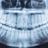
To measure the radiation from dental x-rays on these areas, Dr. Ludlow and his colleagues placed thermoluminescent dosimeter chips at 24 locations in a phantom consisting of a small adult skull and material that simulates soft tissue.
Then the researchers performed a full-mouth intraoral series, four posterior bitewings (a premolar and molar on each side), a panoramic, and lateral and posteroanterior cephalometric images. They repeated each of these 10 times and averaged the dose of radiation for each procedure.
21 deaths in a million
The results? Dental patients, according to this study, are receiving between 0.32 and 4.22 times more radiation than previously estimated. The dose ranged tremendously from one procedure to the next: a bitewing with F-speed film and rectangular collimation exposed the patient to an effective dose (the weighted sum of all organs exposed) of 5 microsieverts (µSv). By contrast, a full-mouth series with D-speed film and round collimation exposed the patient to 388 µSv.
|
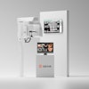
Spare your patients Buying state-of-the-art x-ray equipment may help, but most dentists can significantly reduce their patients' exposure to radiation using the equipment they already have, said Edwin Parks, D.M.D., M.S., director of dental radiology at the Indiana University School of Dentistry in Indianapolis.
|
Dr. Ludlow and his colleagues used a risk coefficient of 0.055 cancer events per sievert to calculate how much harm dental radiation causes. They determined that the risk of fatal cancer ranged from 0.3 in a million for a lateral cephalometric x-ray, to 21 in a million for a full-mouth series with D-speed film and round collimation.
The finding reinforces ADA recommendations (JADA, September 2006, Vol. 137:9, pp. 1304-1312) that dentists should use F-speed film and rectangular collimation, and take x-rays more sparingly, the investigators wrote. The combination of the fast film and rectangular collimation cuts patients' radiation exposure by a factor of 10, they noted.
In an e-mail to DrBicuspid.com, Dr. Ludlow said most dentists are still using the older methods. "The Nationwide Evaluation of X-Ray Trends (NEXT) survey which was published in 2003 found that 73% of dental practices were using D-speed film in 1999. While that figure has been changing, with more practices adopting digital radiographic technologies, the majority of practices continue to use D-speed film. We don't have nationwide data on the use of rectangular collimation, but using North Carolina as a yardstick shows that only 20% of practitioners were using rectangular collimation in a survey completed in 1991," he said (Oral Surgery, Oral Medicine, Oral Pathology, Oral Radiology, and Endodontology, January 1995, Vol. 79:1, pp. 122-126).
The report should spur many dentists to change the way they do radiography, stated Allan Farman, B.D.S., M.B.A., Ph.D., D.Sc., a professor of radiology at the University of Louisville of Kentucky, in an e-mail to DrBicuspid.com. "This is an excellent paper.... Perhaps it is time to ban D-speed film and round collimation for intraoral radiography, and replace these with F-speed film or digital equivalent and with rectangular collimation restricting the beam to the area of the sensor employed. I for one would strongly favor such a move."
He also suggested that U.S. dentists should follow the practice of dentists in Japan and the U.K., who rely more on ultrasound for some procedures.
Edwin T. Parks, D.M.D., M.S., director of dental radiology at the Indiana University School of Dentistry in Indianapolis, said many U.S. dentists are hurting their patients by not keeping up with the latest radiographic procedures.
Dr. Parks said he would favor regulations restricting dentists to rectangular collimation. The change won't happen by itself, he said. "Dentists need to be told."